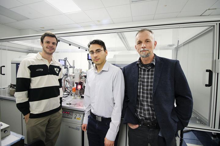
This photo shows SLU researchers Ian Miller, Konstantin Malley and Sergey Korolev, Ph.D.
New insight into a key enzyme associated with various diseases, including Parkinson’s, could lead to new drug candidates.
A team from Saint Louis University (SLU) has found the structure of a key protein that is involved in the body’s inflammatory response, which could pave the way for new treatments of several illnesses, including heart disease, diabetes, cancer, and neurodegenerative diseases like Parkinson’s disease.
The researchers examined the calcium-independent enzyme phospholipase A2β, (iPLA2β)—which cleaves phospholipids in membranes—to better understand how the enzyme is activated during an injury, how it hydrolyses substrates and how it gets shut down, turning the inflammatory response off.
While the enzyme has long been studied, very little is understood about it.
“It was first discovered more than 20 years ago at Washington University in Richard Gross’s lab,” Sergey Korolev, PhD, an associate professor of biochemistry and molecular biology at SLU, said in a statement. “They found that the protein played a role as a part of the cardiovascular system in response to an ischemia or injury.
“Next, researchers found that it is also involved in the insulin production cycle and, when misregulated, can lead to type I diabetes. Then, less than 10 years ago, it was rediscovered from a completely different point of view through the genetic sequencing of patients with neurodegenerative issues. For example, inherited mutations in this gene were identified in patients with early onset Parkinson’s.”
The protein, which is also called PARK14, includes numerous inherited mutations of the gene that was identified in patients with early Parkinson’s.
In the study, the team found that the protein played different roles in different tissues and parts of the cell. The protein’s changeable roles added to difficulties in understanding how it operated.
X-ray crystallography allows the team to gather more data about the protein’s molecular structure by growing a crystal of the protein, shooting x-ray beams through the crystal and analyzing the diffraction pattern generated on a detector plate.
“Before we had the structure, people didn’t have good tools to study this enzyme,” Korolev said. “Now, this will take the field to a new level.
“We’ve opened up lots of possibilities. The mechanism of regulation was completely unknown. Now, the 3D structure gives us a clear hypothesis for how it is responsible for action in different cellular compartments and tissues. Now that we can better understand how the protein interacts with lipid molecules, it will be much easier to develop drugs.”
According to Korolev, the structure they found significantly revised previously developed theoretical models that did not explain many functional features.
“In the past, people have studied this complex enzyme, like a black box, without knowing what is inside,” Korolev said. “Now that we have discovered the structure, we can see every atom. This allows us to visualize what is happening with this protein. It is a completely new level of insight.”
Scientists can now employ different strategies, such as small molecule drugs, to either prevent the enzyme from interacting with other proteins or change its activity to prevent inflammation.
The study was published in Nature Communications.


