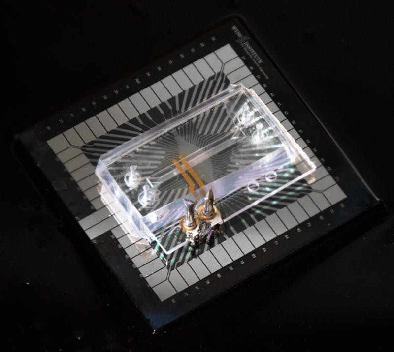
This photograph of the Transepithelial Electrical Resistance- Multi-Electrode Array (TEER-MEA) chip shows the TEER electrodes in gold, the MEA collectors made of platinum in gray, and the two transparent parallel running microfluidic channels on top of the MEA electrodes. Credit: Wyss Institute at Harvard University
Organs-on-Chips (Organ Chips) are emerging as powerful tools that allow researchers to study the physiology of human organs and tissues in ways not possible before. By mimicking normal blood flow, the mechanical microenvironment, and how different tissues physically interface with one another in living organs, they offer a more systematic approach to testing drugs than other in vitro methods that ultimately could help to replace animal testing.
As it can take weeks to grow human cells into intact differentiated and functional tissues within Organ Chips, such as those that mimic the lung and intestine, and researchers seek to understand how drugs, toxins or other perturbations alter tissue structure and function, the team at the Wyss Institute for Biologically Inspired Engineering led by Donald Ingber has been searching for ways to non-invasively monitor the health and maturity of cells cultured within these microfluidic devices over extended times. It has been particularly difficult to measure changes in electrical functions of cells grown within Organ Chips that are normally electrically active, such as neuronal cells in the brain or beating heart cells, both during their differentiation and in response to drugs.
Now, Ingber’s team has collaborated with Wyss Core Faculty member Kit Parker and his group to bring solutions to these problems by fitting Organ Chips with embedded electrodes that enable accurate and continuous monitoring of trans-epithelial electrical resistance (TEER), a broadly used measure of tissue health and differentiation, and real-time assessment of electrical activity of living cells, as demonstrated in a Heart Chip model.
Ingber, M.D., Ph.D., is the Wyss Institute’s Founding Director and also the Judah Folkman Professor of Vascular Biology at HMS and the Vascular Biology Program at Boston Children’s Hospital, as well as Professor of Bioengineering at the Harvard John A. Paulson School of Engineering and Applied Sciences (SEAS). And Parker is also the Tarr Family Professor of Bioengineering and Applied Physics at SEAS.
“These electrically active Organ Chips help to open a window into how living human cells and tissues function within an organ context, without having to enter the human body or even remove the cells from our chips,” said Ingber. “We can now start to study how different tissue barriers are wounded in real time by infection, radiation, drug exposure or even malnutrition, and how and when they heal in response to new regenerative therapeutics.”
The TEER measurement is used to quantify the flow of ions between electrodes and across the tissue-tissue interface made of an organ-specific epithelium and endothelium that is a core component of many of the Institute’s human Organ Chips. Epithelial cells form tissue layers that cover our skin and the inner surfaces of most of our internal organs, while endothelial cells line the adjacent blood-transporting vessels and capillaries that support their functions. Both of these cell layers act as a barrier to small molecules and ions that protects the organs and enables specialized functions, such as absorption in the intestine or urine secretion in the kidney. Conversely, drug toxicities, infections, inflammation and other injurious stimuli can disrupt these barriers. TEER measurements, that are based on restriction of ion passage or electrical resistance, can thus be used to assess both the baseline functional integrity of these cell layers and damage responses that are triggered by drugs or other toxic agents. “Using a new layer-by-layer fabrication process, we created a microfluidic environment in which TEER-measuring electrodes are integral components of the chip architecture and are positioned as close as possible to the tissues grown in one or both of two parallel running channels,” said Olivier Henry, Ph.D., a Wyss Institute Staff Engineer who was the driving force behind the new Organ Chip designs. “In contrast to past electrode designs, this fixed geometry allows accurate measurements that are fully comparable within and between experiments, and that tell us exactly how tissues like that of lung or gut mature within a channel, keep in shape and break down under the influence of drugs or other manipulations.”
The Wyss team’s TEER-measuring Organ Chip design is published in Lab on a Chip. Other authors in addition to Ingber and Henry were Remi Villenave, Ph.D., a Postdoctoral Fellow working with Ingber at the time of the study, and Wyss Researchers Michael Cronce, William Leineweber and Maximilian Benz.
In a second study also reported in Lab on a Chip, the Ingber-Henry team collaborated with Kit Parker who has a strong research interest cardiac biology. Working together, this Wyss interdisciplinary team further enhanced the functionality of the TEER chips by integrating Multi-Electrode-Arrays (MEAs) in the chips that can measure the behavior of electrically active cells like beating heart muscle cells.
Using the TEER-MEA chip, the researchers built a beating vascularized Heart Chip in which human cardiomyocytes are cultured in one microfluidic channel that is separated by a thin semi-permeable membrane from a second, parallel endothelium-lined vascular channel. To test the chip’s new capabilities, the team treated the vascularized Heart Chip with a known inflammatory stimulant that specifically disrupts endothelial barriers or a heart stimulant that acts directly on cardiomyocytes.
“This new chip enables us to perform live electrophysiological measurements to assess the integrity of the endothelial barrier in the heart using TEER measurements, while simultaneously quantifying the beating frequency of the heart cells using MEA. This allows us to reveal how drugs affect heart functions in a scenario where the two cell populations are closely coupled,” said Ben Maoz, Ph.D., a co-first author on the second study, who also is a Technology Development Fellow at the Wyss Institute and a member of Parker’s group. Maoz shared the first-authorship with Henry and Anna Herland, Ph.D., who worked as a Postdoctoral Fellow on Ingber’s team, and is now Assistant Professor at the Royal Institute of Technology and the Karolinska Institute in Stockholm, Sweden.
“The future of Organs-on-Chips is instrumented chips: the idea that the experimenter is taken out of the loop during data collection. Continuous data collection off of organ mimics is what we need to measure efficacy and safety of drugs during long-duration experiments. These kinds of technologies offer us a granularity we have not had before,” said Kit Parker.


