Notorious asphyxiator carbon monoxide has few true admirers, but it’s favored by University of California, Irvine scientists who use it to study other molecules.
With the aid of a scanning tunneling microscope, researchers in UCI’s Center for Chemistry at the Space-Time Limit employed the diatomic compound as a sensor and transducer to probe and image samples, gaining an unprecedented amount of information about their structures, bonds and electrical fields.
The findings were published in Science Advances.
“We used this technique to map, with sub-molecular spatial resolution, the chemical information inside one molecule,” says co-author V. Ara Apkarian, CaSTL director and UCI professor of chemistry.
“To be able to see the inner workings of the basic units of all matter is truly amazing, and it’s one of the main objectives we have pursued at CaSTL for more than a decade.”
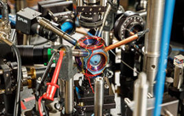
To achieve these results, CaSTL scientists attached a single carbon monoxide molecule to the end of a sharp silver needle inside the scope. They illuminated the tip with a laser and tracked the vibrational frequency of the attached CO bond through the so-called Raman effect, which leads to changes in the color of light scattered from the junction.
The effect is feeble, only one part per billion or so, according to Kumar Wickramasinghe, a UCI professor of electrical engineering & computer science and veteran CaSTL faculty member who was not involved in this study.
But the tip of the needle in the scanning tunneling microscope acts like a lightning rod, amplifying the signal by 12 orders of magnitude.
By recording small changes in the vibrational frequency of the CO bond as it approached targeted molecules, the researchers were able to map out molecular shapes and characteristics due to variations in electric charges within a molecule.

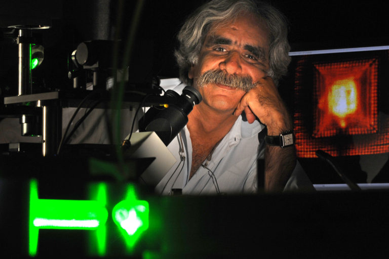
To be able to see the inner workings of the basic units of all matter is truly amazing, and its one of the main objectives we have pursued at CaSTL for more than a decade, says study co-author Ara Apkarian, director of UCIs Center for Chemistry at the Space-Time Limit. Image: Daniel A. Anderson/UCI
The probed molecules in the experiments were metalloporphyrins, compounds found in human blood and plant chlorophyll that are exploited extensively in display technologies.
The captured images provided unprecedented detail about the target metalloporphyrin, including its charge, intramolecular polarization, local photoconductivity, atomically resolved hydrogen bonds and surface electron density waves — the forces that dictate the functionality and structural transformation of molecules. In other words, chemistry.
“Professor Apkarian and his group have, for the first time, created an instrument that can map local electric fields at the sub-molecular level,” says Wickramasinghe, who, as a fellow at IBM, was one of the principal inventors of the world’s earliest atomic force microscope.
“The major step the team has taken is to have made it possible to map the electric field distributions inside a single molecule using the Raman effect, which is a remarkable achievement.”
According to lead author Joonhee Lee, CaSTL research chemist, one of the key results of the experiments was the elucidation of the electrostatic potential surface of the metalloporphyrin molecule — basically, its functional shape, which until recently had been a theoretical construct.
He says the ability to determine this will be particularly beneficial in future studies of macromolecules, such as proteins.
This work is very much in the realm of pure, fundamental science research, Lee notes, but he thinks there may be some practical applications for single-molecule electromechanical systems in the near future.
“Microelectromechanical systems are deployed in current technologies such as smartphones. They take their name from the micron-size scale of such devices; one micron is one-hundredth the size of a human hair,” Lee says.
“Single-molecule electromechanical systems are 10,000 times smaller. Imagine if our miniaturized devices used circuits on that scale.”
The CaSTL project was supported by the National Science Foundation.
Source: University of California, Irvine


