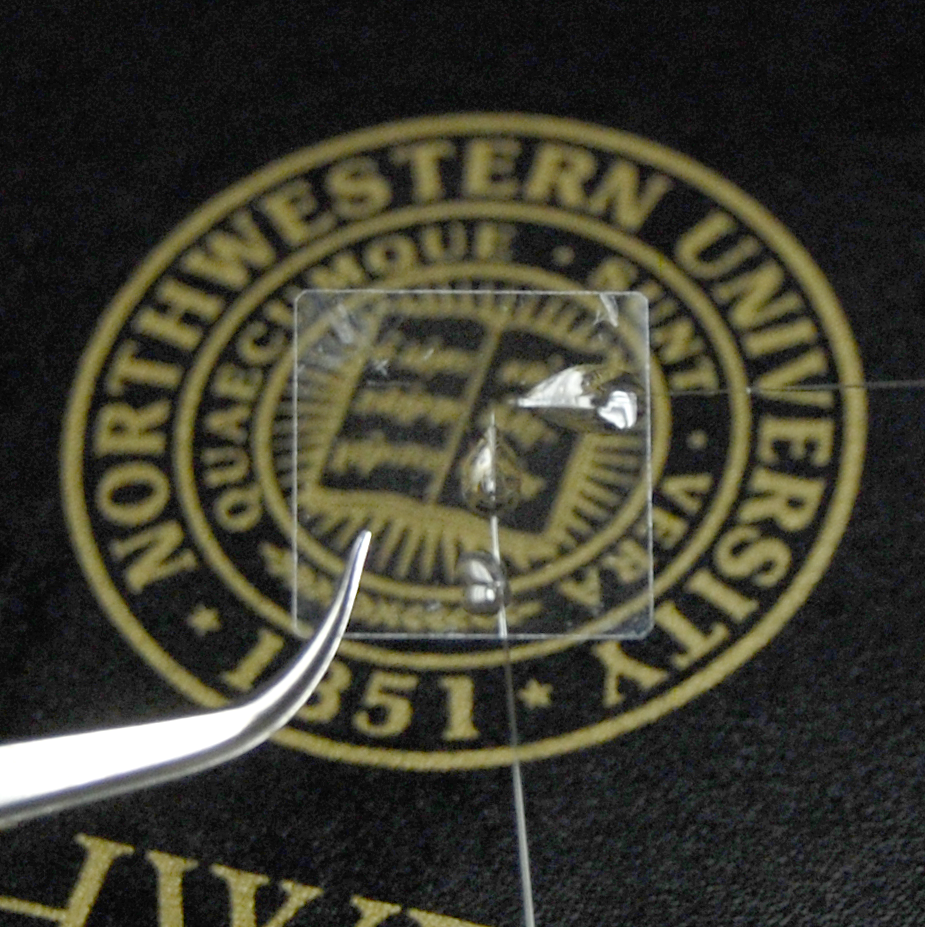
The tiny, transparent Micro-ring device can fit into a contact lens. Credit: Cheng Sun
A new device that is small enough to fit into a contact lens may lead the way for a method to conduct ultrasounds and other scientific applications.
Scientists at Northwestern University have developed a Micro-ring resonator detector, a tiny, transparent device that can help a range of scientific endeavors.
The device can determine the speed of the blood flow and the oxygen metabolic rate at the back of the eye, which could help doctors diagnose common and debilitating diseases like macular degeneration and diabetes.
Other uses may include fundamental biological investigations and clinical diagnosis, from nanoscopic cellular imaging to human breast cancer screening.
The Micro-ring builds on professor Hao Zhang’s 2006 work that combined sound and light waves to create images of biological materials.
Zhang, with a research team, has worked for the last three years to create the device, which is 60 micrometers in diameter and one micron high.
“We believe that with this technology, optical ultrasound detection methods will play an increasingly important role in photoacoustic imaging for the retina and many biomedical applications,” Zhang said in a statement. “We needed a device that had large enough bandwidth for spatial resolution.
“And it needed to be optically transparent to allow light to go through freely.”
According to the study, photoacoustic imaging often uses piezoelectric transducers to detect laser-induced ultrasonic waves.
However, these typically lack adequate broadband sensitivity at ultrasonic frequency higher than 100 MHz, whereas their bulky size and optical methods of ultrasound detection were developed and shown their unique applications in PA imaging.
Cheng Sun, associate professor of mechanical engineering, explained how the size of the device is revolutionary.
“Ultrasound detection devices of that time were usually bulky, opaque and not sensitive enough,” Sun said in a statement. “And they had limited bandwidth.
“It could only capture part of it what was happening in the eye,” he added.
The team plans on improving the device with support from Northwesters, the National Institutes of Health, Argonne National Laboratory and the National Science Foundation. About 12 scientists from various fields have also approached the team about adapting it for their own work.
This includes neuroscientists who have also expressed interest in the device as a way of studying drug protection for the cortex during different points of a stroke in a rodent brain.
“Typically, researchers use a pure piece of glass, but this allows for a lot more types of imaging,” Zhang said.
The study was published in Transactions on Biomedical Engineering.


