Suffering from the resolution limits imposed by diffraction, fluorescent microscopy gets a super-resolution face lift.
 Traditional fluorescence microscopy has suffered from the resolution limits imposed by diffraction and the finite wavelength of light. Classical resolution is typically limited to about 200 nm in xy. Due to the nanoscale architecture of many biological structures, researchers developed super-resolution techniques, starting in the 1990s, to overcome this classical resolution limit in light microscopy. These techniques can be categorized into two regimes: one that treats the sample as a collection of single molecular labels, and a second that views the specimens as a continuously varying density of fluorophores.
The first category is based on the principle of localizing individual fluorescent molecules by Gaussian curve fitting of the emission spots. However, to apply single-molecule localization to a densely labeled specimen in which the emission spots overlap required innovations in imaging that enabled only a subset of the fluorophores to be “switched on” at any given time. One such innovation is called “stochastical optical reconstruction microscopy” (STORM), in which a super-resolution image is constructed by localizing individual molecules in a series of imaging cycles during which only a fraction of fluorophores are switched per cycle. “Using this technique, molecules can be localized to nanometer-scale accuracy and resolution can be improved 10-fold,” says Lynne Chang, senior application scientist, Nikon Instruments Inc.
The second category of super-resolution methods is based on the concept of utilizing spatially non-uniform illumination light to extract sub-resolution information. The technique is called “structured illumination microscopy”, or SIM. In this technique the sample is illuminated with spatially structured light, “which brings normally inaccessible high-resolution information into the observable regime of the microscope in the form of moire fringes,” says Chang. SIM achieves a two-fold improvement in resolution.
Also in the 1990s, Max Planck Institute for Biophysical Chemistry’s Stefan Hell, winner of the Nobel Prize for Chemistry in 2014, realized non-linearity of light-sample interactions can have the potential of theoretically unlimited resolution and developed stimulated emission depletion (STED). Soon after, based on the single-molecule work of William Moerner, Eric Betzig and Harld Hess, from Carl Zeiss, developed the concept of photoactivated localization microscopy (PALM), in which they used photo-switchable proteins to excite only a few single molecules to their bright state among many others remaining in their dark form. “This allows users to precisely localize the singled-out emitters,” says Klaus Weisshart, Carl Zeiss Microscopy. “Since its invention, many improvements and variants of PALM have surfaced.”
Transforming research
Fluorescence microscopy has moved from a technique that’s known for just providing exquisite images to a tool to study cellular structures and processes in a quantitative way. Super-resolution microscopy provides the resolving power needed to distinguish the behavior of single molecules and molecule ensembles from bulk biological samples.
“Likewise, only super-resolution technologies have the power to study interactions and relationships of different molecules in multi-protein assemblies or macromolecular machines under live cell conditions,” says Weisshart. “In addition the abundance, distribution and movement of molecules can be assessed to high precision by particle tracking and counting.”
 Also, now more than ever, scientists are facing pressure to publish their results fast in a quickly evolving scientific world. At the same time, there’s a trend to share high-tech instruments like super-resolution microscopes in multi-user facilities to enhance efficiency. Therefore, according to Isabelle Koester, Leica Microsystems, a super-resolution instrument needs to be easy and convenient to use and show high flexibility.
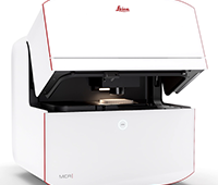
“Time spent on user training, system set-up and maintenance needs to be kept at a minimum,” says Koester. “Combining methods, like STED and fluorescence correlation spectroscopy (FCS), requires flexible systems, like the Leica TCS SP8 confocal platform, which can upgrade any advanced imaging functionality.”
Super-resolution solutions
Five years before any other company entered the super-resolution microscopy market, Leica Microsystems entered the market. The company celebrated its 10th anniversary in super-resolution in 2014 and has won two R&D 100 Awards–in 2012 and 2014–for its super-resolution microscopy products–the Leica SR GSD 3D (2012) and Leica TCS SP8 STED 3X (2014).
The Leica SR GSD 3D technology is based on a technique developed by Stefan Hell called ground state depletion followed by individual molecule return. This method allows users to control the emission of fluorochromes, localizing the position of single molecules far below the diffraction limit. This allows the microscope to achieve an image resolution to about 20 nm.
The Leica TCS SP8 STED 3X, unlike prior systems, which restrict optical resolution improvements to the lateral direction only, extends improvements into the axial direction. Based on the concept of STED, also developed by Hell, the TCS SP8 STED 3X uses superimposed lasers to reduce the effective focal spot scanning the specimen to an area smaller than the diffraction limit.
In 2010, Carl Zeiss entered the super-resolution microscopy market with the ZEISS ELYRA PS.1, the first commercial system to combine the super-resolution techniques of SIM and PALM into one platform. This microscope gave researchers the possibility to choose the technique most appropriate for their samples and to map localized molecules within a super-resolved structural context. Shuttle & Find, introduced in 2012 for the ELYRA PS.1, provided the first correlative workflow between super-resolution and scanning electron microscopy, “thus enabling the mapping of molecules within an ultra-structural context,” says Weisshart.
In 2013, Carl Zeiss launched the 3D PALM measurement up to 2-um sections that can be super-resolved in the axial direction. The 3D PALM received a 2014 R&D 100 Award. The same year also saw the advent of Airyscan, which can be added along with the LSM 880 laser scanning microscope to the ELYRA platform. “Airyscanning, for the first time, provides a confocal super-resolution technique that, similar to the widefield-based SIM, doesn’t require non-linear light-sample interactions,” says Weisshart. As such, it provides excellent sectioning capability and allows live-cell observations at gentle conditions.
 Like Leica Microsystems and Carl Zeiss, Nikon Instruments also provides super-resolution solutions, but focuses on modularity. The company provides solutions in both categories of super-resolution with the N-SIM and N-STORM systems, both marketed in 2009.
“To address the rising need for improved speed for live-cell super-resolution imaging, Nikon has implemented an Overlapping Peak algorithm for N-STORM, which identifies and fits more molecules per imaging cycle, thereby reducing the number of acquisition frames and overall imaging time needed to reconstruct a super-resolution image,” says Chang. The new algorithm also enables more densely labeled samples to be imaged and improves localization density in the reconstructed image.
Nikon has also implemented technology to enable simultaneous two-color N-SIM imaging to drastically improve acquisition times for multi-color SIM imaging. The new feature aids in multi-color live-cell SIM imaging.
Enhancements needed
And while super-resolution microscopy has opened up new discovery opportunities in the biological world, enhancements are still needed to current technology, as no gain is without costs in microscopy. All super-resolution techniques share a common drawback: Spatial resolution can be increased only if temporal resolution is sacrificed.
 In addition, the higher the resolution, the fewer the molecules that will contribute photons, which limits signal-to-noise ratio of the image. Further improvements in imaging speed while maximizing light dosage to cells to prevent phototoxicity will be required to enable super-resolution imaging of highly dynamic events in cells, according to Chang. “Development of new dyes/probes–and corresponding light sources and optical components–will also be necessary to enable imaging of a larger number of molecules,” says Chang.
A super future
Super-resolution microscopy will become a commodity for imaging laboratories globally. Seeing subcellular structure in nanoscopic detail opens a new world to scientists, as it will help to solve cell organelle architecture, the mechanics of molecular motors and the formation and function of macromolecular complexes. Organisms, tissues and cells are 3-D, and multi-color applications give access to detailed information about the interrelationships of these various structures.
Synergies with electron microscopy are expected, according to Weisshart, as correlative microscopy methods provide high-target specificity within an ultra-structural context. Adaptation to light-sheet microscopy will help to further increase live cell compatibility and speed of acquisition.
Finally, super-resolution techniques will leave their marks in optogenetics. “As super-resolution techniques mature, we will witness a significant improvement in our understandings of the inner workings of the cell as the fundamental building of life: live and in 3-D,” says Weisshart.


