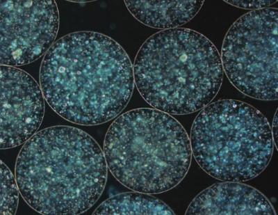
Caption: Researchers have developed a new 3D model that could shed more light on tuberculosis. Credit: The University of Southampton
A new 3D modeling system will help researchers better understand tuberculosis (TB).
A team from the University of Southampton and the University College London have developed an electrostatic encapsulation technique to produce tiny 3D spheres within human cells, which are then infected with TB bacteria to generate conditions that more closely reflect conditions in patients.
This model will allow researchers to further investigate what happens in a human body when TB develops, and may lead to the identification of new antibiotic treatments and vaccines.
Professor Paul Elkington, who leads the Southampton TB research group, explained that the new model will help duplicate patient conditions better than any previous model.
“We believe this is a really exciting development for the field of tuberculosis research,” Elkington said in a statement. “The 3D sphere can be created with a collagen matrix so it is more like a human lung.
“This produces an environment which allows particular antibiotics that are important in treating patients to kill the infection, which they cannot do in other 2D model systems,” he added. “This system will help us speed up the process of finding treatments and vaccines for human tuberculosis, an infection that kills 1.8 million people per year.”
Mycobacterium tuberculosis (Mtb) kills more people annually than any other infection, and is characterized by a spatially organized immune response and extracellular matrix remodeling.
Several other recent clinical trials and vaccine studies to reduce the global burden of tuberculosis have been unsuccessful.
According to the study, antimicrobial resistance is a significant threat to human health because of the emergence of drug-resistant organisms.
The new system incorporates virulent mycobacteria, primary human blood mononuclear cells and collagen-alginate matrix, to dissect the host-pathogen infection.
The 3D spheres can prolong experiments for up to three weeks, more than four times longer than the current standard 2D model systems, which gives researchers more information about how the infection develops the effect of different interventions over time.
The researchers will now collaborate with the African Health Research Institute in Durban, South Africa on a project being funded by a Medical Research Council Global Challenges Research Fund Foundation Award worth $436,170.
Durban has a very high incidence of TB and ideal laboratory infrastructure to introduce the 3D model to study cells from patients at high risk of tuberculosis.
“We are delighted to extend our research and have the opportunity to combine diverse expertise to develop an advanced laboratory system that can be applied to a wide range on infections, especially the infections that are prevalent in resource-poor countries,” Elkington said. “We will use our 3D model to integrate engineering and biological approaches with clinical specimens to create an entirely new system of studying infection.”
Al Leslie, Ph.D., of the Africa Health Research Institute added that the work could lead to further breakthroughs.
“There is a huge amount to be gained from infectious disease biologists and engineers working together, as they push each other out of their comfort zones and force a new perspective on the problem being tackled,” Leslie said in a statement. “This grant is the start of what we hope to be a long-term collaboration that will bring real innovation to our TB research programs and speed up the pace of discovery to fight this deadly epidemic.”
The study appeared in mBio.


