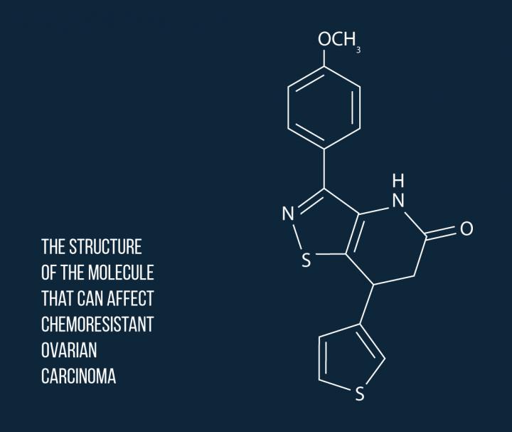
This is the structure of the molecule that can affect chemoresistant ovarian carcinoma. Source: MIPT’s Press Office
A team of scientists from N. D. Zelinsky Institute of Organic Chemistry of the Russian Academy of Sciences (RAS), N. K. Kol’tsov Institute of Developmental Biology of the RAS, and Immune Pharmaceuticals LLC led by MIPT’s Prof. Alexander Kiselyov has synthesized an antitumor compound that could be used to fight chemoresistant cancer. The research findings were published in the European Journal of Medicinal Chemistry.
The scientists synthesized a range of new compounds and evaluated their anticancer effect using sea urchin embryos and human cancer cell lines. One molecule proved to be potent and selective enough to have an effect even on a chemoresistant cancer–ovarian carcinoma. The synthesized compounds belong to a class known as aminoisothiazoles.
“We made the decision to experiment with aminoisothiazoles because compounds of this class exhibit a diverse range of pharmacological and biological activities. This led us to expect that, with the appropriate functional groups, they might act as anticancer agents,” comments Alexander Kiselyov.
The proposed approach enables a straightforward synthesis of the target compounds in high yields. The developed reaction sequence involves only six steps and is based on readily available reagents. To assess antitumor activity, the researchers used in vitro assays based on human cancer cells, as well as the in vivo sea urchin embryo model developed and validated by the team earlier.
Of the synthesized 37 compounds, 12 molecules were found to reduce the proliferation rate of cancer cells with different potencies or to prevent their division completely leading to the death of cancer cells. The effect of these antitumor compounds is attributed to their capacity to destroy microtubules, which are involved in cell division (mitosis). Microtubules are made of a protein called tubulin, which can be targeted by the anticancer agents, causing the degradation of the microtubule structure.
The potency of the synthesized compounds targeting microtubules was further assessed using sea urchin embryos, as well as a panel of human cancer cells from breast adenocarcinoma, melanoma, ovarian and lung tumors. Sea urchin embryos have been shown by the team to be a good model to study specific tubulin-binding agents. They cause embryos to rapidly rotate, as opposed to moving in the ordinary way (cf. the left and right sides of the animation). This effect can be easily observed using a light microscope enabling the scientists to evaluate the anticancer potential of a compound within a short amount of time. Moreover, the team found sea urchin embryos to be more sensitive toward the identified agents than cancer cells. A difference between the duration of the mitotic cycles of sea urchin embryos and cancer cells (40 minutes vs. 24 hours) may lead to distinct effects of the small molecules on tubulin dynamics and could thus account for this phenomenon.The molecule featuring 3-thiophene- and para-methoxyphenyl substituents was identified to be the most potent anticancer agent in the studies. According to the researchers, it is this combination of functional groups, as well as the unique topology of the molecule that are responsible for its unique activity. Specifically, the new agent displays antitubulin properties as it blocks cell division by affecting microtubules and thus can destroy the chemoresistant cells of ovarian carcinoma.
The scientists plan to use crystallography data and structure modeling techniques to study microtubule degradation in more detail in order to identify the sites, or “spots” where the active compounds bind to tubulin.
In their earlier study, the researchers used substances isolated from dill and parsley seeds to synthesize glaziovianin A, another anticancer compound, along with its structural analogs.


