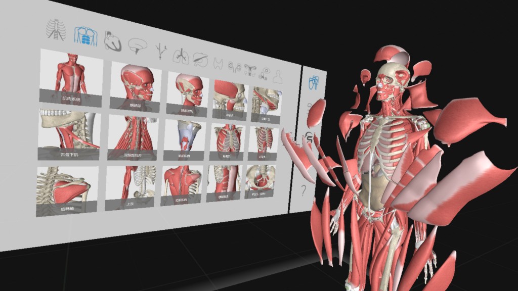
Screenshot of HTC and TMU’s VR anatomy course (HTC image)
Anatomy students at Taipei Medical University are now able to see every internal organ, tissue and muscle in unprecedented 3D detail, thanks to the world’s largest virtual reality (VR) anatomy lab.
The lab, which opened late last year, includes 10 sets of VIVE Pro Headsets loaded with 3D Organon VR Anatomy software, allowing students to both train by themselves and in groups, as multiple users can join a virtual space and experience a human anatomy demonstration.
“With VR providing invaluable course elements, we tutors become navigators to students, who can truly immerse themselves in the virtually-constructed anatomy space as if piloting the best aircraft money can buy,” Hung-Ming Chang, Director of the Department of Anatomy and Cell Biology, Taipei Medical University, said in a statement. “Through virtual reality, we may truly observe the human anatomy in ways and angles that were previously near impossible to delve into. Through a combination of static structure comprehension paired with dynamic representations of spatial human construction, we may greatly boost the understanding and interest of our medical students.”
The new software contains more than 4,000 realistic human body structures, organs and physiological animations. Users can walk around in a virtual environment, while observing different angles of the body, including the skeleton, muscles, tissue, blood vessels, nerves and organs.
Traditionally, researchers have relied on textbooks and 2D models, which are limited because these methods are unable to accurately portray dimensional perceptions and students must visualize how veins, nerves and organs work together within the human body. Cadavers, which can only be used once, are also limited at most medical schools.
Tablet devices and digital anatomy tables have recently been used to study anatomy, but they do not allow the immersive views available in virtual reality, where up to 300 students can study the virtual human body simultaneously.
The new technology also supports dynamic anatomic models that accurately simulate how the heart contracts and the movements of valves in a beating heart muscle.
“VR delivers an accurate visual multi-dimension representation of the human anatomy, allowing for new learning methods that will transform medical education as well as greatly boost its effectiveness,” Edward Chang, HTC President of the Healthcare Division, said in a statement. “We are delighted to see VR applied into mainstream medical education and clinical uses, and hope that this tool will truly benefit more students, tutors, and clinical professionals, as well as the patients themselves.”
Lecturers have already implemented the new technology at the medical school to demonstrate different angles of the body structure. The plan is to combine the VR tools with more traditional education techniques like studying with cadavers.
The university will also be developing VR specific curriculum for students during all stages of matriculation and will look at more applications for the VR tools in the School of Continuing Studies, Advanced Medical EMBA courses, and even into summer camp curriculum for elementary, middle school and high school students.


