Research & Development World

  • R&D World Home
  • Topics
    • Aerospace
    • Automotive
    • Biotech
    • Careers
    • Chemistry
    • Environment
    • Energy
    • Life Science
    • Material Science
    • R&D Management
    • Physics
  • Technology
    • 3D Printing
    • A.I./Robotics
    • Software
    • Battery Technology
    • Controlled Environments
      • Cleanrooms
      • Graphene
      • Lasers
      • Regulations/Standards
      • Sensors
    • Imaging
    • Nanotechnology
    • Scientific Computing
      • Big Data
      • HPC/Supercomputing
      • Informatics
      • Security
    • Semiconductors
  • R&D Market Pulse
  • R&D 100
    • Call for Nominations: The 2025 R&D 100 Awards
    • R&D 100 Awards Event
    • R&D 100 Submissions
    • Winner Archive
    • Explore the 2024 R&D 100 award winners and finalists
  • Resources
    • Research Reports
    • Digital Issues
    • R&D Index
    • Subscribe
    • Video
    • Webinars
  • Global Funding Forecast
  • Top Labs
  • Advertise
  • SUBSCRIBE

Novel Microscopy Technique Developed to Analyze Cellular Focal Adhesion Dynamics

By R&D Editors | June 4, 2018

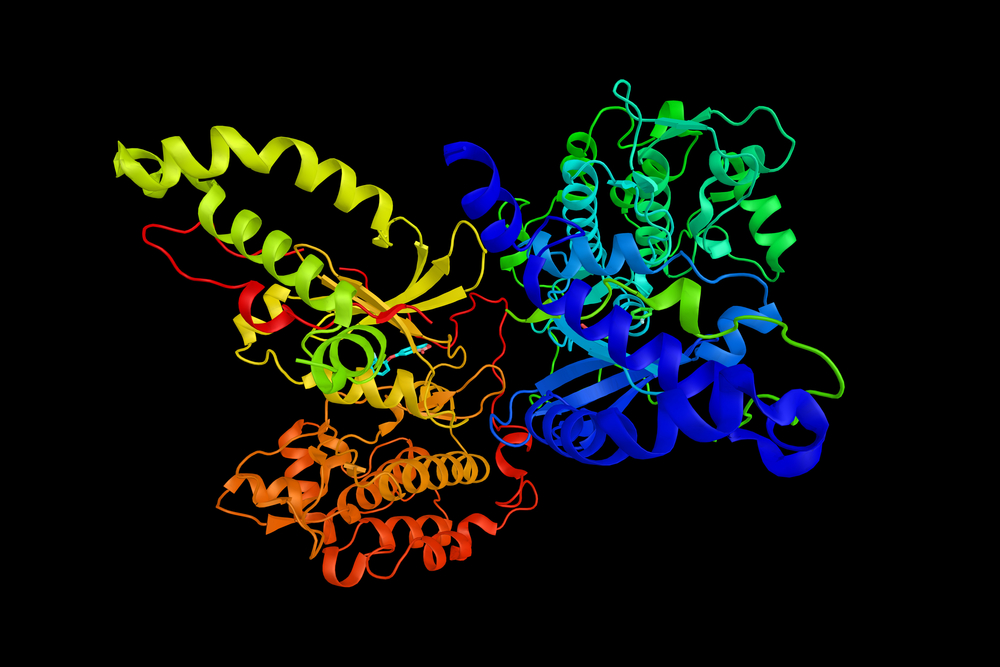
Focal adhesions are large specialized proteins that are located in the area where a cell membrane meets the extracellular matrix (ECM), a collection of molecules surrounding the cells that provide support and regulate micromechanical signals to the cells. Examining focal adhesions is one of the key elements to understanding how a cell proliferates, differentiates, and migrates–which can help in the treatment of diseases like cancer.

Researchers at the Beckman Institute for Advanced Science and Technology and the Micro and Nanotechnology Laboratory at the University of Illinois have developed a new form of microscopy that allows them to observe the formation and evolution of cell membrane focal adhesions. Their paper, “Quantitative Analysis of Focal Adhesion Dynamics Using Photonic Resonator Outcoupler Microscopy (PROM)” details how the new live cell imaging technique can observe the formation and evolution of cell membrane focal adhesions. It was recently published in Light: Science & Applications.

“This is a new kind of biophysics method used to measure the peak intensity shift (PIS) of the spectra reflected from the biomaterials on a photonic crystal surface,” said Yue Zhuo, a Beckman Institute Postdoctoral Fellow and first author on the paper. “The PIS indicates the variation of cluster size in the focal adhesion area of the cell while it’s alive.”

Previous methods involve labeling the cells with fluorescent dyes or tags, which not only can change the physical and chemical makeup of the cell, but also can prove cumbersome for researchers. “Typically people look at focal adhesions with fluorescent tags or proteins,” Zhuo said. “But fluorescent imaging is an invasive imaging method that may change the conformations or block the binding sites of the proteins in the focal adhesion area.”

Fluorescence microscopy is severely limited by an effect called “photobleaching,” in which the fluorophores only maintain their brightness for several seconds. However, many important cell processes occur over the course of minutes, hours, or days. Because PROM does not use fluorophores, and only uses low-intensity illumination, there is not a limit to how long live cells can be measured.

“In the future, we plan to use PROM to study stem cell differentiation, which can occur over the course of several weeks,” said Zhuo.

PROM utilizes a photonic crystal biosensor surface to create an evanescent field (a surface-bound electromagnetic field), which selectively illuminates only the ECM-attached cell membrane and associated protein aggregates directly inside the cell membrane. The photonic crystal strictly limits lateral propagation of light while keeping light tightly bound to the biosensor surface, to enable high-resolution imaging of the cell membrane attachment footprint.

“PROM is providing real-time information about dynamic processes that occur specifically on cell membranes that is not available by any other method,” said Brian Cunningham, a professor of electrical and computer engineering and bioengineering, and the principle investigator for the PROM project. “Since so many biological processes are mediated through attachment of cells to surfaces, PROM provides a unique view of migration, chemotaxis, chemotoxicity, differentiation, biofilm formation, and division. We see PROM as an exciting new tool for cell biologists that can also be applied towards personalized anticancer drug selection, tissue engineering, and sensor-integrated tumor modeling.”

Related Articles Read More >

Eli Lilly facility
9 R&D developments this week: Lilly builds major R&D center, Stratolaunch tests hypersonic craft, IBM chief urges AI R&D funding
professional photo of wooly mammoth in nature --ar 2:1 --personalize sq85hce --v 6.1 Job ID: 47185eaa-b213-4624-8bee-44f9e882feaa
Why science ethicists are sounding skepticism and alarm on ‘de-extinction’
ALAFIA system speeds complex molecular simulations for University of Miami drug research
3d rendered illustration of the anatomy of a cancer cell
Funding flows to obesity, oncology and immunology: 2024 sales data show where science is paying off
rd newsletter
EXPAND YOUR KNOWLEDGE AND STAY CONNECTED
Get the latest info on technologies, trends, and strategies in Research & Development.
RD 25 Power Index

R&D World Digital Issues

Fall 2024 issue

Browse the most current issue of R&D World and back issues in an easy to use high quality format. Clip, share and download with the leading R&D magazine today.

Research & Development World
  • Subscribe to R&D World Magazine
  • Enews Sign Up
  • Contact Us
  • About Us
  • Drug Discovery & Development
  • Pharmaceutical Processing
  • Global Funding Forecast

Copyright © 2025 WTWH Media LLC. All Rights Reserved. The material on this site may not be reproduced, distributed, transmitted, cached or otherwise used, except with the prior written permission of WTWH Media
Privacy Policy | Advertising | About Us

Search R&D World

  • R&D World Home
  • Topics
    • Aerospace
    • Automotive
    • Biotech
    • Careers
    • Chemistry
    • Environment
    • Energy
    • Life Science
    • Material Science
    • R&D Management
    • Physics
  • Technology
    • 3D Printing
    • A.I./Robotics
    • Software
    • Battery Technology
    • Controlled Environments
      • Cleanrooms
      • Graphene
      • Lasers
      • Regulations/Standards
      • Sensors
    • Imaging
    • Nanotechnology
    • Scientific Computing
      • Big Data
      • HPC/Supercomputing
      • Informatics
      • Security
    • Semiconductors
  • R&D Market Pulse
  • R&D 100
    • Call for Nominations: The 2025 R&D 100 Awards
    • R&D 100 Awards Event
    • R&D 100 Submissions
    • Winner Archive
    • Explore the 2024 R&D 100 award winners and finalists
  • Resources
    • Research Reports
    • Digital Issues
    • R&D Index
    • Subscribe
    • Video
    • Webinars
  • Global Funding Forecast
  • Top Labs
  • Advertise
  • SUBSCRIBE