A Danish research team is behind a new method for studying how a tracer is distributed in a cancer tumor via its extensive vascular network.
The method can be used for purposes such as closely studying the effect of medical treatment using cancer inhibitors.
By means of mathematical modelling, the researchers combined two previously known scanning technologies – magnetic resonance imaging (MRI) and computed tomography (CT) – and used these to study tumors in laboratory animals.
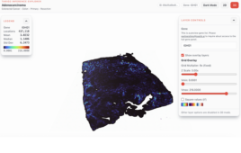
This resulted in completely new images at very high resolution, which provide detailed mapping of the branching of tumor blood vessels.
“We can lay two images of the same cancer tumor on top of each other so to speak, so we get a more geometrically complex understanding of the individual tumor’s blood vessels, and thereby an opportunity to very precisely study the way drugs are distributed,” says Associate Professor Jens Vinge Nygaard, Department of Engineering, Aarhus University.
He is responsible for the mathematical modelling work for the imaging, and he expects that the method could ultimately be used to develop new drugs and optimise dosing for the individual patient.
15,000 blood vessels under the microscope
An MR image can show how a tracer used as a cancer-inhibiting drug is distributed inside the tumor, but only in a relatively coarse resolution.
An image from a micro-CT scanner, on the other hand, can show an extensive network of blood vessels in the tumor at very high resolution, but it is unable to identify how the drug is transported locally.
The combination of the two imaging technologies can thereby provide significantly improved scanning images of cancer, which can play an important role in developing new drugs.
“The new images give us an opportunity to follow the way a tracer travels through the blood vessels in the tumor and into the surrounding tissue to the cancer cells. As scientists, we’re interested in mapping the size and branching of the blood vessels, and understanding what goes on between the blood vessels over time. This can provide us with more detailed insight into specific treatment needs,” says Thomas Rea Wittenborn, who is a cancer researcher at the Department of Experimental Clinical Oncology, Department of Clinical Medicine, Aarhus University Hospital.
The tumors they studied measure approximately 200-300 cubic millimetres and typically contain 15,000-20,000 branches of blood vessels.
Computer models replace experimental animals
The researchers followed a total of ten mice with tumors on their feet, and used the two scanning technologies to develop a computer model for each of these.
In principle, the models form the foundation for a completely unique experimental platform.
“Using the computer models, we’ve created a virtual experimental platform so to speak, and can thereby considerably extend our experiments because we’re not dependent on experimental animals. In practice, we can sit in front of our computer screens and study what happens to the tumor if we use drugs that stay in the tissue for longer or shorter periods, or are adapted for small or large blood vessels,” says Associate Professor Nygaard.
In the time ahead, the researchers will expand their experiment with more mice and follow the cancer tumors over a period of time. This will provide them with an opportunity to develop computer models that not only describe drug distribution in a static stage of the cancer process, but also generate precise growth scenarios for cancer tumors.
“Using the new imaging method, we’ll be capable in purely mathematical terms of predicting tumor development in connection with different drug strategies,” says Associate Professor Nygaard.