This schematic depicts an imaging system that uses a special type of laser beam called a Bessel beam that is produced using a pair of cone-shaped “axicon” lenses combined with a microscope objective. Purdue University researchers are using the system, which is able to penetrate deep into tissue and might lead to technologies that eliminate the need to draw blood for analyses including drug testing and early detection of diseases such as cancer and diabetes. Image: Purdue University photo/Ji-Xin Cheng
A “chemical imaging” system that uses a special type of laser beam to penetrate deep into tissue might lead to technologies that eliminate the need to draw blood for analyses including drug testing and early detection of diseases such as cancer and diabetes.
The system, called stimulated Raman projection microscopy and tomography, makes possible “volumetric imaging” without using fluorescent dyes that might affect biological functions and hinder accuracy, said Ji-Xin Cheng, a professor in Purdue University’s Weldon School of Biomedical Engineering, Department of Chemistry and Birck Nanotechnology Center.
“Volumetric chemical imaging allows a better understanding of the chemical composition of three-dimensional complex biological systems such as cells,” he said.
The technology uses a type of laser beam called a Bessel beam, which maintains focus for a longer distance than a traditional “Gaussian beam” used in other imaging technologies, making it possible to penetrate deep into tissue. Stimulated Raman spectroscopy eliminates the need for fluorescent dyes. The technology yields more accurate data than other methods because it allows imaging of the entire cell by “adding up” signals produced from the scanning beam, Cheng said.
Because the Bessel beam makes possible deep-tissue imaging, it could lead to systems that eliminate the need to draw blood for analyses such as drug testing and detection of biomarkers for non-invasive early diagnosis of diseases, Cheng said.
“This is a long-term goal,” he said. “In the meantime, much more research is needed to improve the system.”
The researchers proved the concept by imaging fat storage in living cells. Findings are detailed in a research paper appearing on April 24 in the journal Nature Communications.
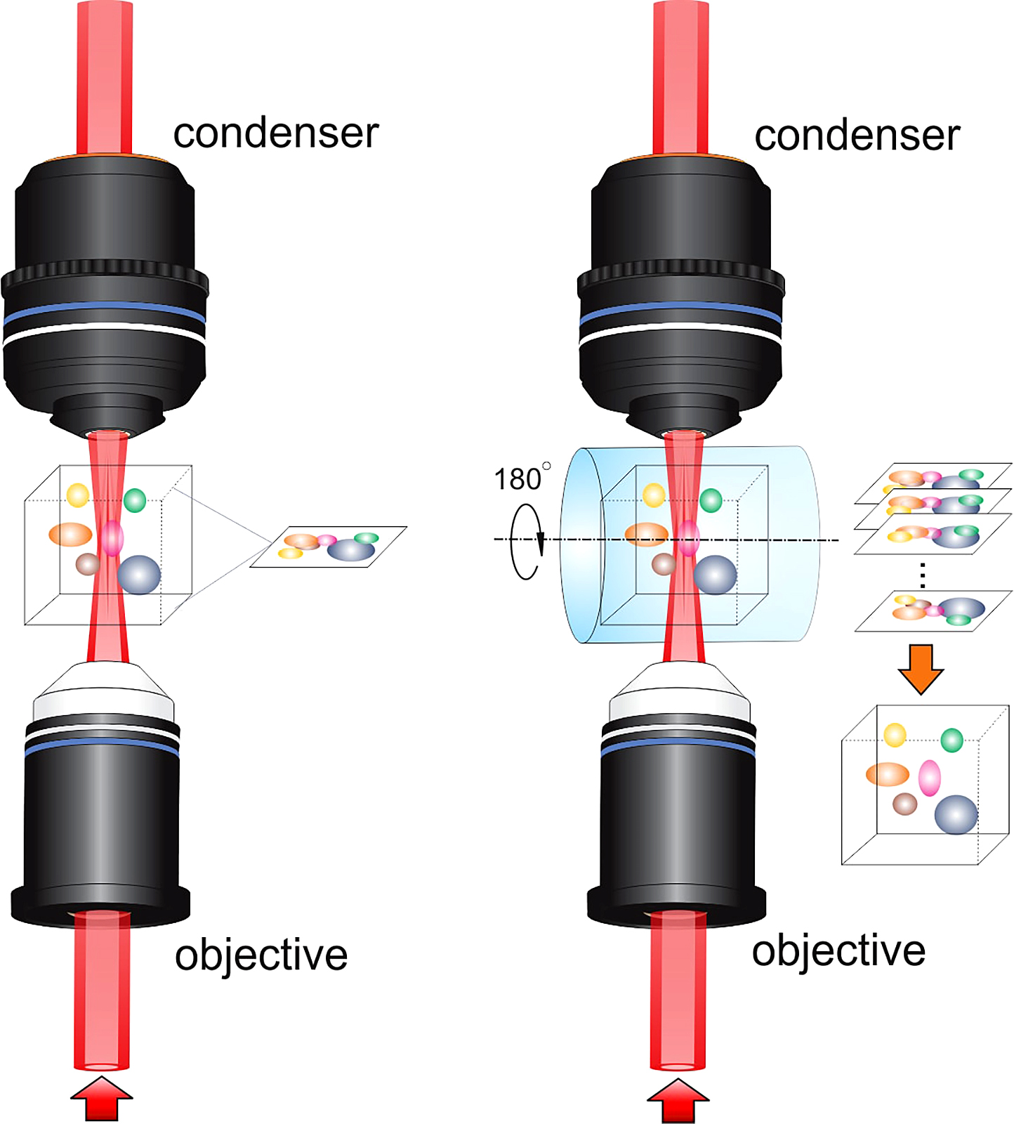
The reported technology yields information about chemical composition, collecting a series of images while rotating the sample and reconstructing the 3-D structure through image reconstruction algorithms.
The Bessel beam is produced using a pair of cone-shaped “axicon” lenses and is combined with a microscope objective. Its use for volumetric fluorescence imaging was previously demonstrated by physicist Eric Betzig, who won the Nobel Prize in chemistry in 2014 for his pioneering contribution to super-resolution fluorescence microscopy. Super-resolution technology allows researchers to resolve structural features far smaller than the wavelength of visible light, sidestepping the “diffraction limit” that normally prevents imaging of features smaller than about 250 nanometers, which is large compared to certain biological molecules and structures in cells.
However, fluorescence microscopy usually requires the use of fluorescent tags, which may interfere with biological processes and hinder accuracy for determining chemical structure.
Future research will include work to increase the detection sensitivity of the system and improve the imaging quality and speed.
“There is plenty of room for improvement,” Cheng said. “The system is based on a bulky and relatively expensive femtosecond laser, which limits its potential for broad use and clinical translation. Nevertheless, we anticipate that this limitation can be circumvented through engineering innovations to reduce the cost and size of our technology. We also note that the Bessel beam can be produced using fibers, which could simplify the system and enable endoscopic applications.”
The paper was authored by Xueli Chen, a visiting scholar from Xidian University in China; Purdue postdoctoral research associate Chi Zhang; Purdue doctoral students Peng Lin and Kai-Chih Huang; Xidian University researchers Jimin Liang and Jie Tian; and Cheng.
The research was supported by funds from the Keck Foundation and National Institutes of Health.
Source: Purdue University