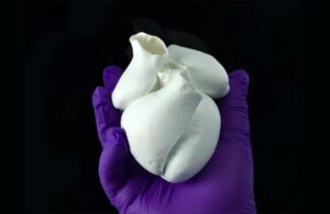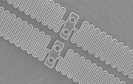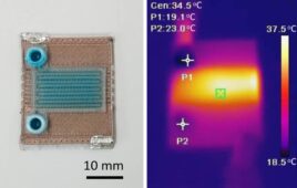
A biohybrid model of a four-chambered heart engineered with focused rotary jet spinning (FRJS) technology [Image courtesy of Harvard SEAS]
Harvard University researchers used focused rotary jet spinning (FRJS) technology to fabricate polymer fibers that mimic the helical structure of heart muscles.
They created ventricle structures with the method and then seeded them with rat cardiomyocyte or human stem cell-derived cardiomyocyte cells, according to a news release from the Wyss Institute for Biologically Inspired Engineering at Harvard University. Roughly a week later, they had several thin layers of beating tissue covering the scaffold. The cells in the beating tissue followed the same helical alignment as the fibers underneath.
The bioengineers, who were from the Wyss Institue and the Harvard John A. Paulson School of Engineering and Applied Sciences (SEAS), were then able to run experiments that compared the performance of ventricles made from helically aligned fibers with those made from circumferentially aligned fibers. They found the helically aligned tissue outperformed the circumferentially aligned tissue on every front. The result provided evidence backing a decades-old hypothesis — biomathematician Edward Sallin’s idea from 1969 that the heart’s helical alignment is critical to achieving large ejection fractions.
“In this case, we went back to address a never tested observation about the helical structure of the laminar architecture of the heart. Fortunately, Professor Sallin published a theoretical prediction more than a half century ago, and we were able to build a new manufacturing platform that enabled us to test his hypothesis and address this centuries-old question,” said Kit Parker, professor of bioengineering and applied physics at SEAS, an associate faculty member at the Wyss Institute and senior author of the research paper appearing in Science.


