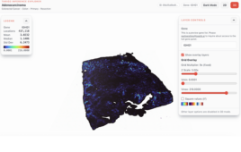
X-ray image of a 2D native electrophoretic separation. The sulfur content, often part of cysteine and methionine residues in proteins, is shown in red. The zinc content is shown in green. By sampling protein that co-localizes with the metal for mass spectrometry, researchers can identify the proteins that bind them. |

Metals such as copper, zinc, and iron are important nutrients to all
life. The special properties of these elements that make them so useful in
technologies including batteries and catalysts—for example, having multiple
stable oxidations states under ambient conditions—also make them useful to living
organisms.
With over a third of all proteins thought to bind metals, knowing which
metals are bound and how that binding changes in response to the environment
could have big implications.
For instance, the biological mismanagement of metals is involved in many
diseases, including Lou Gehrig’s disease, Wilson and Menkes disease, and possibly
even Alzheimer’s disease.
Metals are also an environmental toxin, such as the hexavalent chromium
featured in the movie Erin Brockovich, and they are used in drugs, like
the platinum in cisplatin that treats prostate cancer.
Developing an approach for making determinations about the relationship
of metals and proteins is complex because the experimental methods that are
routinely used to identify proteins, for example denaturing gel
electrophoresis, can also remove metals that might be bound to them.
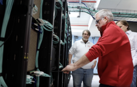
Scientists working at the U.S. Department of Energy Office of Science’s
Advanced Photon Source (APS) at Argonne National Laboratory have made great
strides in imaging metals within cells. Using the X-ray imaging capabilities
afforded by the APS, researchers have seen, often for the first time, where the
metals reside inside cells and tissues. These capabilities have allowed
researchers to see how the elemental content of bacteria change upon adhesion,
fluxes of zinc in egg cells upon fertilization, and changes in the locations
where copper is stored in a cell during the growth of blood vessels.
But many of the images that have been acquired led to new questions: Are
these metals required for the activity of proteins? Which proteins are binding
with which metals inside the cell?
Now, a team of researchers from the Worcester Polytechnic Institute and
Argonne carrying out research at the APS have developed a new experimental
approach that not only detects and distinguishes metals in proteins, but also
characterizes the proteins that bind the metals, without removing them. This
work, which was featured in Metallomics, used X-ray fluorescence imaging
(XRF) at X-ray Science Division (XSD) beamline 8-BM-B of the APS.
|

Employing modified native 2D gel electrophoresis, the researchers were
able to separate proteins from the organisms S. oneidensis, a bacterium
that can reduce poisonous heavy metal and can live in both environments with or
without oxygen, and P. aeruginosa, a common bacterium that can cause
disease in animals, including humans, and that is found in soil, water, skin
flora, and most man-made environments throughout the world. Then, using XRF,
the team quantitatively measured the amount of sulfur, iron, and zinc at every
point of the 2D separation, pinpointed the location of proteins that had metals
bound to them, and determined the identity of these proteins utilizing mass
spectrometry.
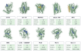
The approach enabled the research team to identify a novel protein
(PA5217) as a zinc-binding protein in P. aeruginosa.
Their finding highlights how this method not only determines changes in
metal occupancy, but also identifies the associated protein.
Now that this new technique is developed, questions raised by images of
the metals in cells can be studied further.
Native 2D gel electrophoresis separation is accessible to most
laboratories, and resources for 2-D XRF imaging are available at the APS.
This development will help researchers begin to identify which of the
one-third of proteins that are thought to bind metals actually do, and what
roles they play in life.
Source: Argonne National Laboratory