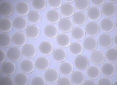
A structure-switching nanosensor made from DNA (blue and purple) detects a specific transcription factor (green). Using these nanosensors, a team of researchers from UCSB has demonstrated the detection of transcription factors directly in cellular extracts. The researchers believe that their strategies will allow biologists to monitor the activity of thousands of transcription factors, leading to a better understanding of the mechanisms underlying cell division and development. Image: Peter Allen |

Sensors made from custom DNA molecules could be used to
personalize cancer treatments and monitor the quality of stem cells, according
to an international team of researchers led by scientists at the University of California,
Santa Barbara and the University of Rome Tor
Vergata.
The new nanosensors can quickly detect a broad class of
proteins called transcription factors, which serve as the master control
switches of life. The research is described in an article published in Journal of the American Chemical Society.
“The fate of our cells is controlled by thousands of
different proteins, called transcription factors,” says Alexis
Vallée-Bélisle, a postdoctoral researcher in UCSB’s Department of Chemistry and
Biochemistry, who led the study. “The role of these proteins is to read
the genome and translate it into instructions for the synthesis of the various
molecules that compose and control the cell. Transcription factors act a little
bit like the ‘settings’ of our cells, just like the settings on our phones or computers.
What our sensors do is read those settings.”
When scientists take stem cells and turn them into
specialized cells, they do so by changing the levels of a few transcription
factors, he explained. This process is called cell reprogramming. “Our sensors
monitor transcription factor activities, and could be used to make sure that
stem cells have been properly reprogrammed,” says Vallée-Bélisle.
“They could also be used to determine which transcription factors are
activated or repressed in a patient’s cancer cells, thus enabling physicians to
use the right combination of drugs for each patient.”
Andrew
Bonham, a postdoctoral scholar at UCSB and co-first author of the study,
explained that many labs have invented ways to read transcription factors;
however, this team’s approach is very quick and convenient. “In most labs,
researchers spend hours extracting the proteins from cells before analyzing
them,” says Bonham. “With the new sensors, we just mash the cells up,
put the sensors in, and measure the level of fluorescence of the sample.”
This international research effort––organized by senior
authors Kevin Plaxco, professor in UCSB’s Department of Chemistry and
Biochemistry, and Francesco Ricci, professor at the University of Rome,
Tor Vergata––started when Ricci realized that all of the information necessary
to detect transcription factor activities is already encrypted in the human
genome, and could be used to build sensors. “Upon activation, these
thousands of different transcription factors bind to their own specific target
DNA sequence,” says Ricci. “We use these sequences as a starting
point to build our new nanosensors.”
The key breakthrough underlying this new technology came
from studies of the natural biosensors inside cells. “All creatures, from
bacteria to humans, monitor their environments using ‘biomolecular switches’––shape-changing
molecules made from RNA or proteins,” says Plaxco. “For example, in
our sinuses, there are millions of receptor proteins that detect different odor
molecules by switching from an ‘off state’ to an ‘on state.’ The beauty of
these switches is that they are small enough to operate inside a cell, and
specific enough to work in the very complex environments found there.”
Inspired by the efficiency of these natural nanosensors, the
research group teamed with Norbert Reich, also a professor in UCSB’s Department
of Chemistry and Biochemistry, to build synthetic switching nanosensors using
DNA, rather than proteins or RNA.
Specifically, the team re-engineered three naturally occurring
DNA sequences, each recognizing a different transcription factor, into
molecular switches that become fluorescent when they bind to their intended
targets. Using these nanometer-scale sensors, the researchers could determine
transcription factor activity directly in cellular extracts by simply measuring
their fluorescence level.
The
researchers believe that this strategy will ultimately allow biologists to
monitor the activation of thousands of transcription factors, leading to a
better understanding of the mechanisms underlying cell division and
development. “Alternatively, since these nanosensors work directly in
biological samples, we also believe that they could be used to screen and test
new drugs that could, for example, inhibit transcription-factor binding
activity responsible for the growth of tumor cells,” says Plaxco.