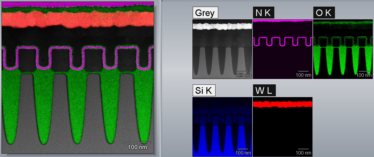
Extended MHN filter, 7X7 kernel, 1 pass; overlaid on gray reference image
The 1960s and 1970s proved to be a revolutionary period for the development of new digital image processing techniques. These efforts were mainly focused on medical imaging at the light microscope level and were developed primarily for gray level images acquired with video cameras.
With the ensuing emergence of solid state and color cameras, many of these techniques were expanded to include digital-color images. Among these techniques is digital image filtering, used to remove bright or black spots or speckles, which are mostly due to random noise (including shot noise and Johnson-Nyquist noise). There have been numerous digital image filters introduced over the long history of image processing. Most of these filters are linear convolution filters, but recently, a series of non-linear smoothing filters has been proposed. These image filters are used to achieve specific image enhancements and are categorized by the results they produce.
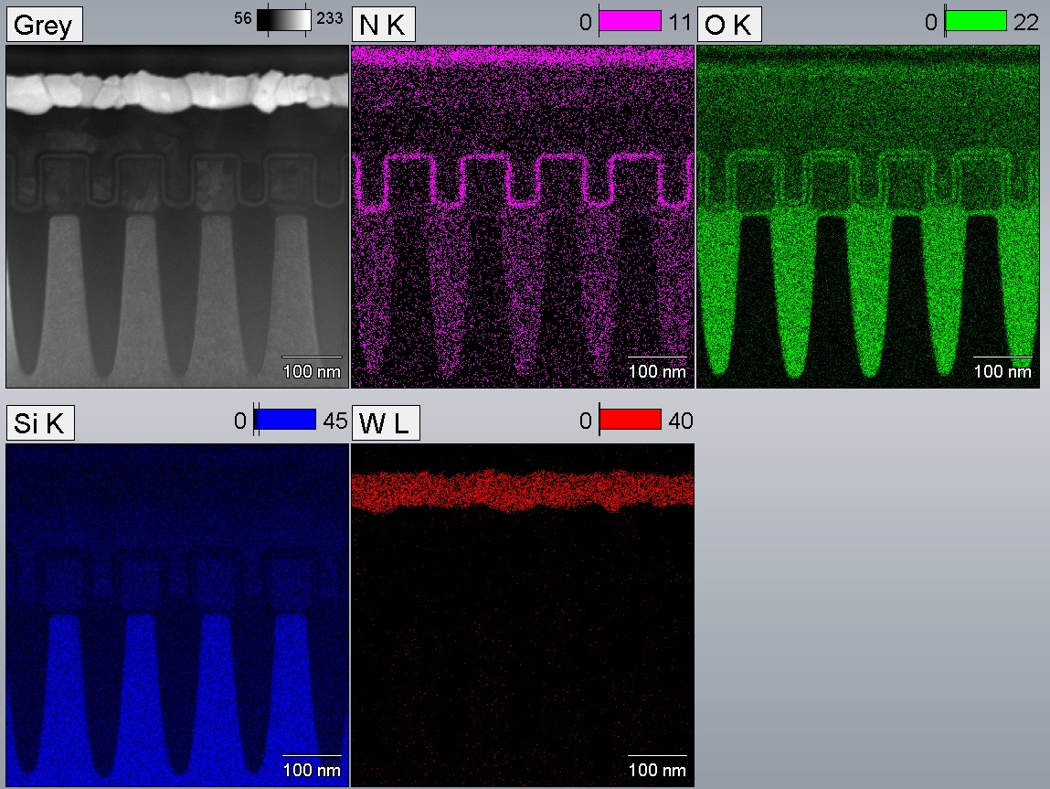
Elemental X-ray images acquired by energy dispersive spectroscopy (EDS) in an electron microscope suffer from some of the same types of noise challenges as digital camera images. These challenges are enhanced when EDS X-ray maps are acquired under less than optimal conditions, such as low accelerating voltages, low beam currents or when elements are in low concentrations and/or distributed in isolated areas within the sample. A few common issues include sparse data where the element maps have only a few (typically less than 10) X-ray counts per pixel, and hot pixel effects, by which a very small number (even one) of pixels has an anomalously high intensity that results in the overall brightness being diminished as the contrast is stretched to capture the hot pixel within the image intensity range.
Applying the algorithms and techniques developed for digital image filtering to X-ray element maps effectively remedies many of the challenges described. Smoothing filters using a large kernel improves the effective statistics of collected X-rays and makes the data easier to interpret by reducing noise and “cleaning up” the speckling of X-ray maps. Edge-finding and sharpening filters mitigate the impact of feature broadening or blurring that may result from application of only a smoothing filter. These same filters also highlight very small features, drawing the eye to otherwise overlooked regions of interest. Hot pixel corrections ensure maximum contrast within the vast majority of the image with minimal saturation of the hot pixels.
The implementation of non-linear, digital X-ray filters offers the advantage of combining feature benefits such as smoothing and sharpening in a single filter. As an example, the extended maximum homogeneity neighbor filter (Extended MHN), which has the dual benefit of acting as a smoothing filter that also preserves edges, is one in a group of maximum homogeneity neighbor filters that check the homogeneity of small areas (often 3X3 pixels within a 5X5 mask) within the close neighborhood of the pixel in consideration.
By applying the latest software algorithms and non-linear image filters, imaging scientists can enhance filter X-ray element maps to improve the collected statistics of X-rays and make data easier to interpret.
The extended MHN filter is shown in here. This sample suffers from sparse data with features on the nanoscale and a pixel resolution of 0.25 nm, making a general smoothing filter not applicable. (Credit: Thermo Fisher Scientific).


