Researchers led by Pacific Northwest National Laboratory have combined two X-ray techniques to watch how a molecule’s electrons and its surrounding water reorganize together during a reaction that underpins photosynthesis and cellular metabolism in what the team says is the first study to combine these techniques to capture that interplay in a single experiment.
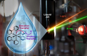
Illustration of the light-induced electron and proton transfer in the PCET reaction, alongside a photo of the liquid jet setup where light and X-ray beams intersect the ruthenium polypyridyl complex.
Credit: Illustration by Jeff London | Pacific Northwest National Laboratory
They published their work in Nature Communications.
The reaction is proton-coupled electron transfer (PCET), which describes any reaction where an electron and a proton both move as part of the same transformation.
During the PCET part of photosynthesis, light knocks an electron off a chlorophyll complex, creating a powerful oxidant that pulls an electron from a nearby amino acid. Simultaneously, the amino acid passes a proton to a neighboring histidine, balancing its charge. Cellular metabolism uses the same mechanism in the opposite direction to take electrons out of sugar, fats and proteins.
Scientists have been studying this mechanism for decades, but no one has been able to capture it in a single study with local and structural sensitivity. Advanced X-ray methods available at the Linac Coherent Light Source at SLAC National Accelerator Laboratory allowed the researchers to do exactly that.
The researchers used ultrafast X-ray spectroscopy, scattering and advanced simulations to capture the key steps in the reaction, establishing a framework that could eventually resolve questions about the order in which the particles move.
The team used a well-studied synthetic ruthenium-based molecule that absorbs light and captures a proton from its surroundings in acidic conditions. They chose the molecule because it does not undergo additional electronic or structural rearrangements that would complicate the signals. The electron transfer finished faster than the 180-femtosecond resolution of the measurement, while the proton took roughly 460 picoseconds to arrive, a gap of more than a thousandfold that let the researchers isolate each step.
The researchers used two instruments in the SLAC laboratory to conduct their work: chemRIXS for element-specific X-ray absorption spectroscopy and the X-ray Correlation Spectroscopy (XCS) instrument for time-resolved X-ray scattering. Together, these methods revealed how electrons moved between molecular sites and, in combination with molecular dynamics simulations, how solvent molecules rearranged.
While the team was able to observe the local reorganization of the electronic structure and water networks, they could not directly observe the proton. This is because X-ray scattering sees atoms that are rich with electrons.
“Understanding the photochemistry of this complex required us to push the limits of our data analysis. By combining X-ray absorption spectroscopy with precise theoretical modeling, we gained an unprecedented look into the real-time electronic changes driving these reactions,” Abdullah Kahraman, the first author of the paper, said in a press release.


Tell Us What You Think!
You must be logged in to post a comment.