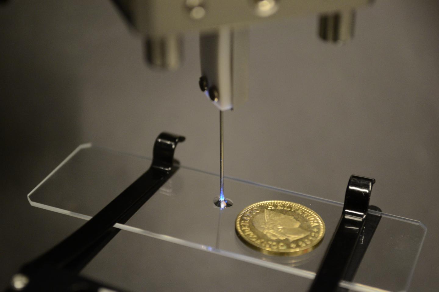
Researchers used an optical fiber housed inside the needle pictured to deliver light for 3-D printing microstructures. The light selectively hardens volumes inside the droplet of photopolymer on the glass slide. The new system could one day allow 3-D printing inside the body. Credit: Damien Loterie and Paul Delrot, École Polytechnique Fédérale de Lausanne
Ultra-thin optical fibers could make it possible to use 3D printers to repair damaged tissues in humans.
A team of researchers have developed an optical fiber as thin as a human hair that can be used to create microscopic structures with laser-based 3D printing and an endoscope to fabric tiny biocompatible structures directly into tissue inside the body.
“With further development our technique could enable endoscopic microfabrication tools that would be valuable during surgery,” research team leader Paul Delrot, from École Polytechnique Fédérale de Lausanne, Switzerland, said in a statement. “These tools could be used to print micro- or nano-scale 3D structures that facilitate the adhesion and growth of cells to create engineered tissue that restores damaged tissues.”
During the study, the researchers created microstructures with a 1.0-micron lateral and a 21.5-micron axial printing resolution, an approach that could be useful for studying how cells interact with various microstructures in animal models.
To create the microstructures, the researchers dipped the end of an optical fiber into a liquid known as photopolymer that solidifies, or cures, when illuminated with a specific color of light. The optical fiber was used to deliver and digitally focused laser light point-by-point into the liquid to build a 3D microstructure.
By printing delicate details onto large parts, the new ultra-compact microfabrication tool could also be a useful add-on to today’s commercially available 3D printers, which are used for everything from rapid prototyping to making personalized medical devices.
“By using one printer head with a low resolution for the bulk parts and our device as a secondary printer head for the fine details, multi-resolution additive manufacturing could be achieved,” Delrot said.
Current laser-based microfabrication techniques rely on a non-linear optical phenomenon called two-photon photopolymerization to selectively cure a volume deep inside a liquid photosensitive material.
However, because two-photon photopolymerization requires complex and expensive lasers that emit very short pulses, as well as bulky optical systems to deliver light, they are difficult to use for biomedical applications.
“Our group has expertise in manipulating and shaping light through optical fibers, which led us to think that microstructures could be printed with a compact system,” Delrot said. “In addition, to make the system more affordable, we took advantage of a photopolymer with a nonlinear dose response.
“This can work with a simple continuous-wave laser, so expensive pulsed lasers were not required.”
The researchers took advantage of a chemical phenomenon where solidification only occurs above a certain threshold in light intensity to selectively cure a specific volume of material and discovered the best parameters to print microstructures using a low-power, inexpensive laser that emits continuously.
The team used an organic polymer precursor doped with photoinitiator made of off-the-shelf chemical components to create hollow and solid microstructures.
They focused a continuous-wave laser emitting light at 488-nanometer wavelength—visible-wavelength light that is potentially safe for cells—through an optical fiber small enough to fit in a syringe.
The researchers focused the light inside the photopolymer so that only a small 3D point was cured. Performing a calibration step prior to microfabrication allowed them to digitally focus and scan laser light through the ultra-thin optical fiber without moving the fiber.
“Compared to two-photon photopolymerization state-of-the-art systems, our device has a coarser printing resolution, however, it is potentially sufficient to study cellular interactions and does not require bulky optical systems nor expensive pulsed lasers,” Delrot said. “Since our approach doesn’t require complex optical components, it could be adapted to use with current endoscopic systems.”
The study was published in Optics Express.