 A team of French and Swiss scientists has demonstrated a lensless imaging technique that could easily be implemented in cost-effective and compact devices in phage laboratories to accelerate phage-therapy diagnosis.
The growing number of drug-resistant bacterial infections worldwide is driving renewed interest in phage therapy. The WHO has warned of “a slow tsunami” of antibiotic resistance that by 2050 could result in 10 million annual deaths from antibiotic-resistant infections.
Based on the use of a personalized cocktail composed of highly specific bacterial viruses, phage therapy employs bacteriophages, a form of virus, to treat pathogenic bacterial infections. Following promising phage-therapy clinical studies treating infection of burn wounds, urinary tract infections and other problems caused by antibiotic-resistant bacteria, a growing body of evidence has built a consensus among scientists that there is synergism between phages and antibiotics.
First implemented in 1919, phage therapy relies on a range of tests on agar media to determine the most active phage on a given bacterial target, or to isolate new lytic phages from an environmental sample. However, these culture-based techniques must be interpreted through direct visual detection of plaques.

In a recent research article, “Phage Susceptibility Testing and Infectious Titer Determination Through Wide-Field Lensless Monitoring of Phage Plaque Growth”, the team reported a lensless technique for testing the susceptibility of the bacterium to the phage on agar and measuring infectious titer, among other results. In addition to CEA-Leti, the team included a Grenoble consortium of researchers from CEA-Interdisciplinary Research Institute of Grenoble (CEA-Irig), the French National Centre for Scientific Research-Laboratoire des Technologies de la Microélectronique (CNRS-LTM), and the phage-therapy team from Lausanne University Hospital in Switzerland, better known as CHUV in French.
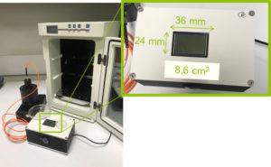
In addition to investigating computer-assisted methods to ease and accelerate diagnosis in phage therapy, the team studied phage plaque using a custom-designed, wide-field lensless imaging device, which allows continuous monitoring over a very-large-area sensor (8.64 cm2).
“We report bacterial susceptibility to anti-Staphylococcus aureus phage in three hours and estimation of infectious titer in eight hours and 20 minutes,” the paper explains. “These are much shorter time-to-results than the 12-to-24 hours traditionally needed, since naked eye observation and counting of phage plaques is still the most widely used technique for susceptibility testing prior to phage therapy. Moreover, the continuous monitoring of the samples enables the study of plaque-growth kinetics, which enables a deeper understanding of the interaction between phage and bacteria.”
With 4.3 μm resolution in the lensless demonstrator, the scientists also detected phage-resistant bacterial microcolonies of Klebsiella pneumoniae inside the boundaries of phage plaques.
“This shows that our prototype is also a suitable device to track phage resistance,” said Pierre Marcoux, a scientist in CEA-Leti’s Department of Microtechnologies for Biology and Health. “Lensless imaging is therefore an all-in-one method that could easily be implemented in cost-effective and compact devices in phage laboratories to help with phage-therapy diagnosis.”


Tell Us What You Think!