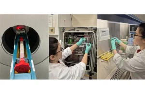
The bioanalytic system, featuring user-friendly assembly, holds reconstructed 3D cell tissue for PET scanning. Precisely regulated conditions within the system allow for accurate analysis of drug distribution and mode of action in a tissue-like environment.
Historically, animal testing has been something of a necessary evil in drug discovery, but a growing number of efforts are underway to do without them. The latest case in point is a newly patented system that integrates 3D cell cultures. These cultures grow human cells on supportive silk scaffolds. This setup better mimics real body tissue. The scaffolds are placed in a special device. It uses controlled fluid flow, much like chromatography systems. This allows automated, real-time PET/CT scans. Researchers at the University of Vienna highlight the potential of analyzing potential cancer imaging agents, radiopharmaceuticals, within this tissue-like environment to do away with the need for animal models.
A ‘unique bioanalytic hybrid system’
Published in The Journal of Nuclear Medicine, the research introduces what the authors call a “unique bioanalytic hybrid system.” That is, it merges advanced cell culture techniques with established analytical methods. In particular, it uses “silk fibroin sponges… to simulate an extracellular matrix,” allowing cells to grow in a realistic 3D “tissue analog.” This scaffold is then placed within a “chromatographic-like system array” that provides continuous, controlled fluid flow. The researchers state this combination harnesses “the strength of chromatographic systems,” enabling automation and high-throughput processing, “with the complexity of an advanced 3-dimensional cell culture” needed to model “real tissue-like geometry, morphology, and dynamics.”
This Austrian research, developed by a consortium including the University of Vienna, Medical University of Vienna, Technikum Wien, and company partner DOC Medikus GmbH, builds on prior preclinical testing research aimed at creating more physiologically relevant in vitro models. Scientists have long recognized that standard 2D cell cultures often fail to predict in vivo responses. In turn, that leads to the development of 3D cultures such as spheroids or cells on scaffolds and dynamic bioreactor systems that mimic tissue structure and fluid flow. Earlier work, such as that by Whitehead and Shoghi cited in the 2013 JNM paper, even demonstrated the feasibility of PET-compatible bioreactors for imaging biomarkers. Yet the novelty presented here lies in the specific “bioanalytic hybrid” integration: using biocompatible silk sponges explicitly as a stationary phase within a chromatographic-like dynamic system designed for direct, automated analysis with small-animal PET/CT. While previous systems explored dynamic cultures or PET imaging in vitro, this work taps chromatographic principles for potentially higher throughput and standardization. It also supports the ability to assess key radiotracer parameters like nondisplaceable binding to the tissue analog alongside specific cellular uptake. It thus aims to bridge the translational gap more effectively for radiopharmaceutical development.
Potential for clinical use
Ultimately, this proof-of-concept study demonstrates a “versatile and simple” bioanalytic system, as its authors note in JNM. Validation experiments confirmed consistent cellular behavior compared to traditional models. The research also adds the capability to gauge parameters like nondisplaceable binding, effectively distinguishing promising radiotracer candidates from poor ones. By aligning with both the 3Rs principles and the FDA’s Critical Path Initiative, this automated, high-throughput approach holds significant potential in curbing the reliance on animal testing. The system features particularly user-friendly assembly and precisely regulated conditions within the apparatus. First author Verena Pichler emphasizes that their method aims to make development of new radioactive marker substances more efficient while improving ethical standards. They also note that it promises to “accelerate and make radiotracer development more accessible,” potentially speeding the “rapid translation of more innovative radiotracers into clinical use.”