Credit: Lindsay Jawor/R&D
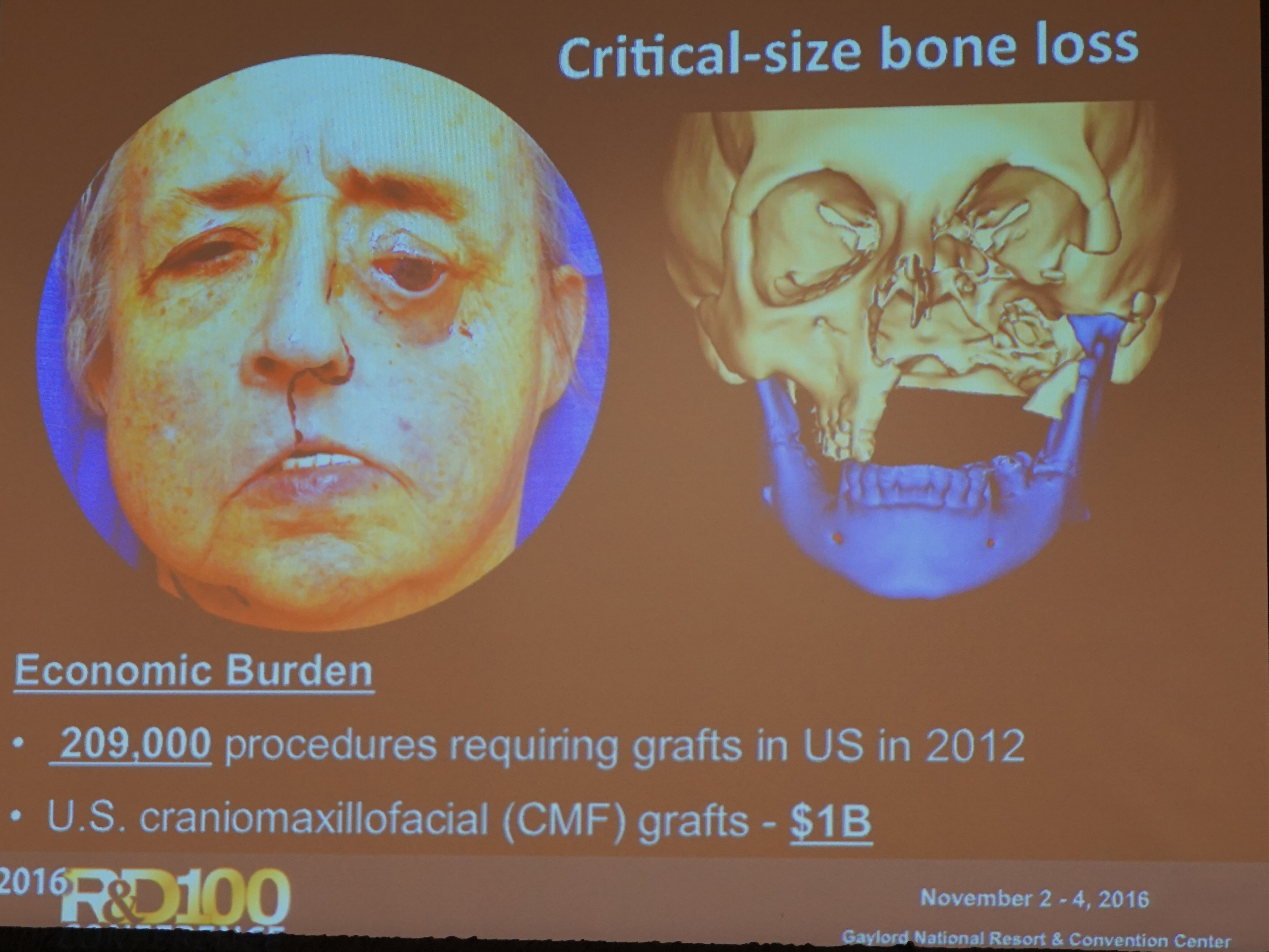
Bone regeneration is a critical need in the United States. Each year, 209,000 procedures require craniomaxillofacial grafts for patients undergoing facial reconstructive surgery.
Currently the sole option surgeons have is to take another bone, break it and fit it into a patient’s facial structure, which is not the ideal solution.
In comes Prof. Warren Grayson, Ph.D., and his team who have created a ready-to-implant plastic bone that can turn into living tissue and dramatically improve life for patients.
“Can we regenerate bone with appropriate anatomical shape and vascular supply?” Grayson asked during his presentation at the second annual R&D 100 Conference that took place earlier this month at the Gaylord National Resort & Convention Center in Washington, D.C.
The answer is yes. In his presentation titled “3D Printing and the Search for Better Bone Replacement,” Grayson spoke about breakthrough technologies that allow for precise control of the cellular microenvironment and addressed fundamental questions regarding the application of biophysical cues to regulate stem cell differentiation.
The Associate Professor in the Department of Biomedical Engineering & Translational Tissue Engineering Center at Johns Hopkins University School of Medicine and his team at the Grayson Lab have developed a successful combination for 3D bone printing by creating an effective framework for filling in missing bone. They have achieved this goal by mixing at least 30 percent of pulverized natural bone with special man-made plastic, which then they molded into the necessary shape using a 3D printer.
While the world of 3D bioprinting is still very new and ambiguous, a few scientists have started utilizing this cutting-edge technology for various areas of regenerative medicine to help fill the tissue-and-organ shortage void.
In their experiments on mice, Grayson and his team decided to make a composite material that would combine the strength and printability of plastic with the biological “information” contained in natural bone. The team held several experiments before finding a successful combination.
The scaffolds were tested on rodents with relatively large holes in their skulls where bones were input experimentally. Without intervention, the bone wounds were too large to heal. Mice that got scaffold implants laden with stem cells saw new bone growth within the hole over the 12 weeks of the experiment. CT scans showed that at least 50 percent more bone grew in scaffolds containing 30 or 70 percent bone powder, compared to those with pure Polycaprolactone (PCL- a biodegradable polyester).
Grayson’s work represents a significant step forward in the 3D printing of human bones.
The Grayson Lab also conducts research on stem cell biology, bioreactor technology and scaffold development.
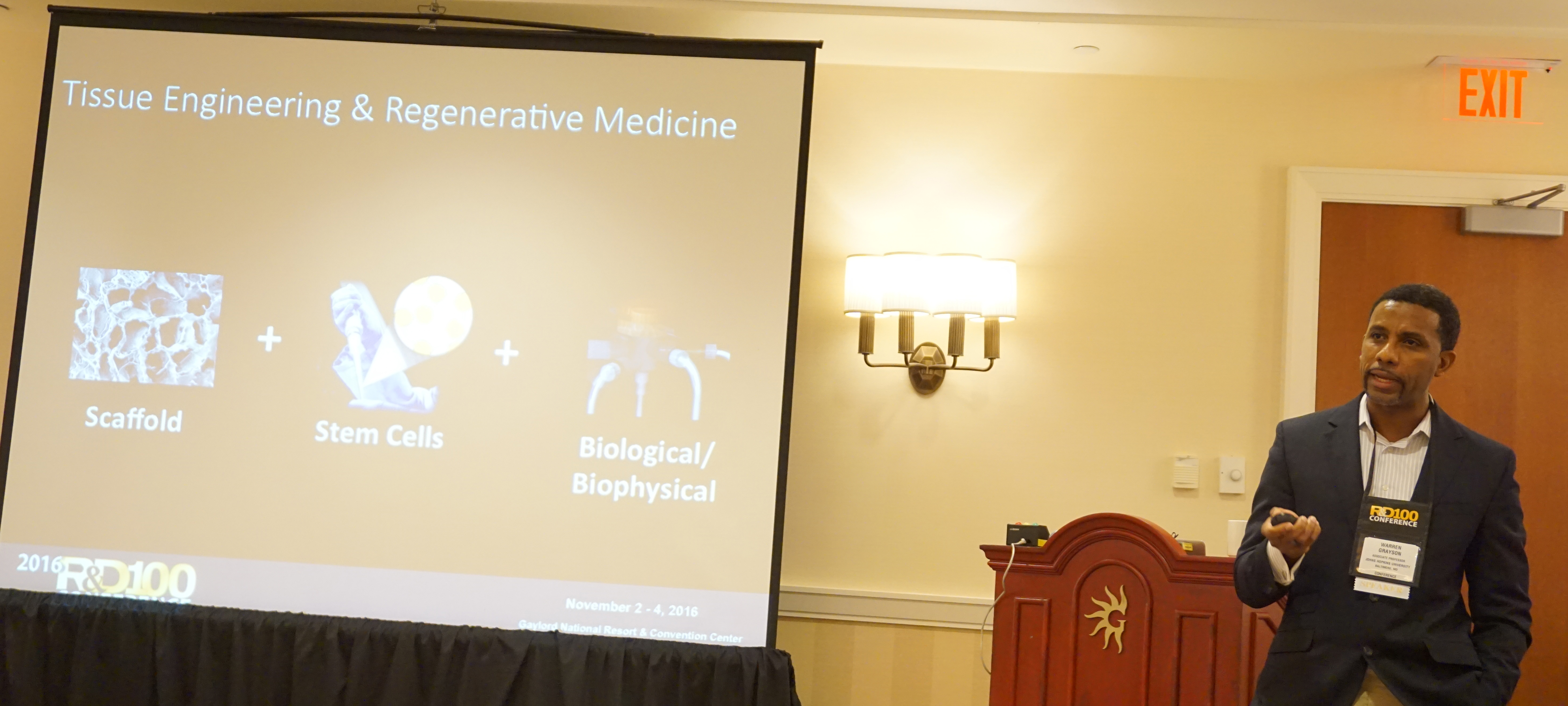
Warren Grayson, Ph.D., talks about better bone replacement at the second annual R&D 100 Conference in Washington, D.C. (Credit: Lindsay Jawor/R&D)
To submit your new product or invention to be considered for the 2017 R&D 100 Awards, please click here.