Scientists from the Robotic Materials Department at the Max Planck Institute for Intelligent Systems in Germany published a paper in Science Advances last week describing biohybrid microrobots that can deliver cancer treatments directly to tumor sites.

Conceptual schematics depicting cell-based microrobots magnetically guided to a target tumor site, where continuous, local TRAIL secretion eliminates cancer cells without affecting nearby healthy tissues. Created in BioRender. N. O. Dogan (2026), https://biorender.com/e18h681. DOI: 10.1126/sciadv.aea9831
Medical microrobots have potential for targeted therapeutic delivery. However, current systems achieve only physical targeting, and once at the target site, they are unable to distinguish healthy cells from cancerous ones because of the lack of biological selectivity.
The biohybrid microrobots are derived from human embryonic kidney cells genetically engineered to produce tumor necrosis factor-related apoptosis-inducing ligand (TRAIL), a molecule that induces cancer cell death in multiple tumor types without damaging healthy cells. TRAIL binds to death receptors 4 and 5, which are overexpressed in cancer cells.
The engineered cells were then conjugated to biocompatible magnetic Janus particles, silica beads half-coated with FePt nanofilms, to enable external magnetic control. With magnetic fields, the microrobots accumulated around tumor spheroids and continuously released TRAIL for several days, leading to selective cancer cell death while avoiding damage to healthy cells.
Engineering for TRAIL secretion and remote control
The researchers optimized the genetic modification of the cells using a nonliposomal transfection reagent and plasmid DNA encoding both TRAIL and Green Fluorescent Protein (GFP). Peak TRAIL secretion, 211 ng/ml, and high transfection efficiency, approximately 96.6%, were achieved at 48 hours post-transfection. These levels were achieved using a 2:1 transfection reagent-to-DNA ratio.
To enable remote control of the cells, they were conjugated with magnetic Janus particles. The particles were 500 nm diameter silica beads, half-coated with a 60 nm thick iron platinum magnetic nanofilm. FePt was selected for its strong magnetic properties, high biocompatibility and high contrast in medical imaging modalities. The particles were half-coated to create the magnetic anisotropy necessary to generate torque, enabling steering of the cells.
The microrobots respond to external rotating magnetic fields, achieving a maximum speed of 107 μm/s at 60 Hz.
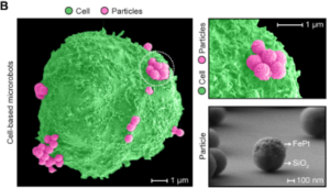
SEM images showing cell-based microrobots with magnetic Janus particles attached to the cell surface, with no evidence of internalization. Images were pseudocolored. Scale bars, 1 μm. SEM image of magnetic Janus particle, confirming the deposition of FePt on its silica core. Scale bar, 100 nm. DOI: 10.1126/sciadv.aea9831
Microrobots eliminate up to 98% of cancer cells
Conditioned medium from the microrobots containing approximately 220 ng/ml of TRAIL was applied to various cell lines, resulting in reductions in cancer cell viability. In glioblastoma, only 2% viability remained. Healthy cell lines showed high resistance to the therapy. BJ human fibroblasts showed a negligible reduction in viability.
The study demonstrated that the microrobot swarms could be navigated toward 3D spheroids using magnetic fields and localized TRAIL secretion effectively induced cell death throughout the tumor structure.
After the microrobots have finished secreting TRAIL, the cells are eliminated by the immune system. The Janus particles are then absorbed by the liver and spleen and degraded. The researchers emphasize that further study is needed to map the long-term clearance of the particles and monitor for accumulation.
The researchers successfully applied the same technology to human skin fibroblasts. They also developed a doxycycline-inducible system, allowing on-demand secretion of TRAIL only after the robots have reached the tumor.
The study also confirmed that the Janus particles remained stably attached to the cell membrane even under high-pressure shear stresses found in human arteries and veins.