The 10x Genomics Xenium Analyzer can slash analysis time from hours to minutes. [10x Genomics]
Today, genomics platform company 10x Genomics is taking its own giant steps in cellular analysis. To date, the company, which was founded in 2012, has developed a trio of platforms that push past conventional limitations: Chromium, Visium, and Xenium.
While many organizations are content with incremental progress, 10x Genomics, as founding scientist Michael Schnall-Levin explains, thrives on tackling “10x problems.” This ethos stems from advice co-founder Ben Hindson received during a stint at Lawrence Livermore National Labs, where a mentor urged him to pursue problems where he could make a tenfold difference. As Hindson, 10x’s chief scientific officer, explained in a 2022 Biomarker interview, “When someone says it’s impossible, that’s when I get interested.”
Schnall-Levin, who has been with the company since its early days, noted that scientific research is inherently demanding. “Even incremental things are frequently kind of hard, so there’s a lot of value in focusing on things that are going to make a big difference, because you’re going to wind up spending a lot of work on it either way,” he said. He adds that the company’s vision is to “develop impactful technologies” that enable scientists to “understand and manipulate biological complexity in meaningful ways.”
Chromium streamlines single-cell workflows
The first of the company’s products, the Chromium platform, enables precise single-cell analysis through microfluidic partitioning, where cells move through 50–60 micrometer channels at limiting dilution. It uses microfluidics to generate nanoliter-scale droplets called GEMs (Gel Beads-in-emulsion). Each GEM contains a single cell, a barcoded gel bead coated with oligonucleotides, and enzymes and reagents for molecular capture.
The Chromium X Series, powered by GEM-X technology, uses flow-based emulsion to GEMs more efficiently than previous iterations. The system can process up to 20,000 cells per channel in under six minutes, with improved cell recovery rates up to 80%.

Michael Schnall Levin, Ph.D.
“The machine combines these in very small microfluidic channels making millions of little droplets where each droplet gets a cell and molecular machinery to copy RNA, proteins, or epigenetic information,” Schnall-Levin explained. The system generates tens of thousands of these nanoscale reaction chambers within minutes.
In October 2024, the company announced GEM-X Flex and GEM-X Universal Multiplex products, which can now analyze cells for less than a penny each, with the ability to profile up to 2.5 million cells per run. “These launches are an integral part of our ongoing push to democratize single-cell analysis,” Schnall-Levin noted in a press release.
The increased throughput and reduced cost of these new products enables researchers to generate larger datasets that can be visualized using established techniques like UMAP (Uniform Manifold Approximation and Projection). The dimensionality reduction technique helps preserve both local and global structures in high-dimensional data, allowing researchers to identify distinct cellular populations and their relationships.

Chromium X
The granular view provided by 10x Genomics’ technology has redefined our understanding of diseases like cancer, where the technology reveals not just tumor heterogeneity but the complex dynamics of immune cells in the tumor microenvironment. As Schnall-Levin explained during the facility tour, “People are looking at CAR-T therapies, which is a big thing now. One of the biggest challenges is that after these engineered T cells are injected into a patient to kill cancer cells, many of them become inactive after a few weeks.” This has been a central limitation in CAR-T therapy efficacy, but single-cell analysis is helping researchers understand why.
This challenge of CAR-T cell inactivation has spurred researchers to seek answers, and single-cell analysis is emerging as a powerful tool. “Researchers can use our products to track these CAR-T cells over time,” Schnall-Levin said. “They can take blood draws from patients who’ve received the therapy and, using single-cell analysis, look back in time to see which T cells continued to attack the cancer and which ones stopped. They can get a molecular understanding of why some T cells remain active while others don’t.”
The power of the analysis lies in its comprehensive nature. “You can tell from the molecular state by looking at the genes that are turned on in a T cell,” Schnall-Levin noted. “We measure around 18,000 genes per cell. There’s enough known biology already that you can look at which genes are expressed at what levels and classify the T cells into different states—whether they’re active, dormant, or even suppressing the immune response.”
Visium preserves spatial context in gene expression
“Spatial biology is fascinating because it combines imaging data with molecular data. With spatial technology, we can overlay detailed molecular data onto tissue images.”
—Michael Schnall-Levin, 10x Genomics
The spatial transcriptomics platform Visium takes a fundamentally different approach by preserving the spatial context of gene expression within tissues. It centers on specialized slides manufactured with high-density arrays of spatially barcoded capture probes. When tissue sections are placed on these slides, RNA molecules are captured by location-specific probes, maintaining their original spatial relationships.
The Visium platform’s slides contain specialized regions called Capture Areas designed to interface with tissue samples. Each standard capture area (6.5mm x 6.5mm) features approximately 5,000 precisely positioned spots, with each spot measuring 55 μm in diameter and spaced 100 μm apart. The specialized oligonucleotide-based technology ensures each spot contains unique spatial barcodes, enabling precise tracking of gene expression.
The latest version, Visium HD, provides unprecedented spatial resolution with 2 × 2 μm squares arranged in a continuous array. This high-definition view has revealed previously hidden aspects of disease biology—from subtle features like tumor borders and small blood vessels to the unexpected mixing of malignant cell states within seemingly uniform tumors.
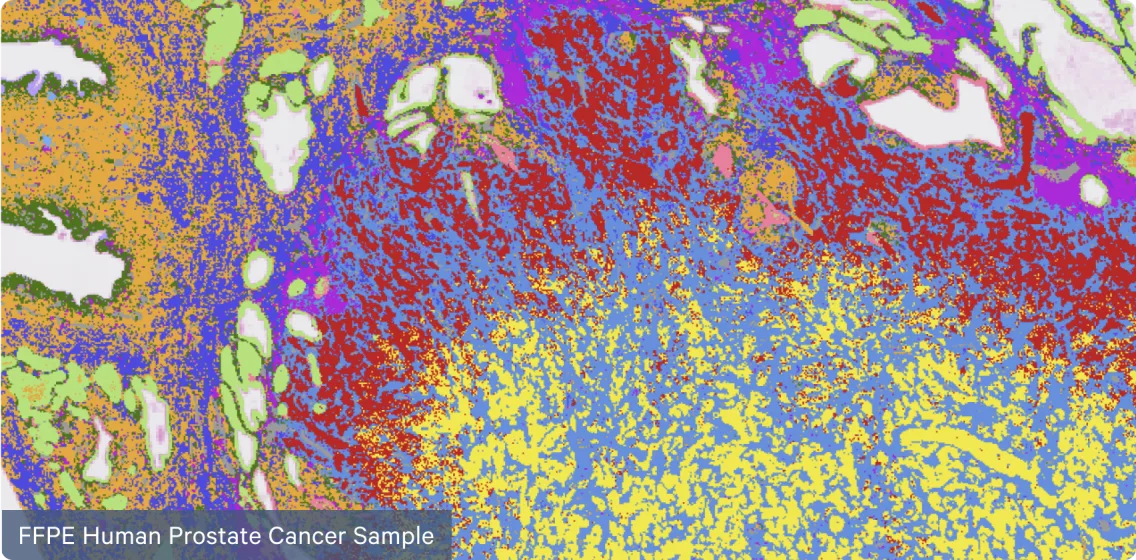
Formalin-fixed, paraffin-embedded (FFPE) human prostate cancer sample shown via Visium HD. [10x Genomics]
In Alzheimer’s disease studies, for instance, the technology has identified specific gene expression changes near amyloid plaques, shedding light on inflammation and myelination. The platform supports both fresh-frozen and FFPE (Formalin-Fixed Paraffin-Embedded) tissue sections, making it a fit for clinical research using archived tissue samples.
Xenium’s subcellular resolution unveils biological complexity
The Xenium platform enables direct visualization of RNA molecules within intact tissue at subcellular resolution. Initially launched in December 2023, the platform expanded its capabilities with the release of the Xenium Prime 5K Pan-Tissue and Pathways panel in June 2024, allowing analysis of up to 5,000 genes simultaneously while processing up to 472 square millimeters of tissue in under six days.
The platform combines automated fluidics for cycling chemical reactions with high-resolution imaging capabilities, achieving single-cell resolution with XY-localization under 50 nanometers and Z-localization below 100 nanometers. “Inside here, there’s advanced imaging happening, mixing in and out cycles of chemicals, and taking pictures at really high resolution of all the molecules inside,” explains Schnall-Levin while demonstrating the instrument. The system supports both fresh-frozen and FFPE tissue samples.
A notable feature of the Xenium platform is its integrated data processing capabilities. “There’s a powerful computer integrated with the instrument, equipped with GPUs to handle the intensive image processing and data analysis,” notes Schnall-Levin. “All the algorithm code is open source on these products, which makes it easier for people to see what’s going on under the hood. All the file formats that the software packages put out are well-documented, usually very standard file formats.”
“We saw some companies trying to make file formats opaque to lock customers into their systems, or trying to build something like an App Store, which hasn’t worked out well,” Schnall-Levin added. “We learned these lessons and focused on interoperability and practicality. Customers have really appreciated that there’s no big cloud uploads and downloads to be able to actually access and process.”
The visualization system enables detailed analysis of molecular expression within tissue contexts. “The display shows individual RNA molecules within the tissue,” said Holly Ross, associate director, technical training, in a demo. “The dots represent detected molecules, and the background image is a staining of the tissue, highlighting structures like cell membranes. The software uses this information to segment the tissue and identify individual cells.”
“This technology allows us to molecularly determine what cells are doing,” Schnall-Levin said. “For example, rather than a pathologist noting a region has many immune cells, we can identify which are attacking and which are suppressing immune responses. Combining these insights is extremely exciting, and we’re just at the beginning.”
“From the beginning of the company, we really wanted software and data informatics to be a core part. Historically in biotech, a lot of companies start with some technology in the lab and then tack on software at the end. We took a different approach.”
—Michael Schnall-Levin, 10x Genomics
Xenium has helped advance researchers’ understanding of complex biological systems. In brain tumor research, the platform revealed previously unrecognized patterns of cellular interaction within glioblastomas. Researchers identified distinct spatial relationships between tumor-associated macrophages and hypoxic tumor cells.
In breast cancer studies, the technology’s ability to simultaneously analyze multiple molecular markers led to the identification of rare cell populations that traditional pathology might overlook, especially in regions where tumors show heterogeneous expression patterns. The platform’s multi-modal analysis capabilities allow researchers to examine RNA expression, protein detection, and morphological features simultaneously on the same tissue section.
“For example, in lung cancer research, scientists mapped the spatial distribution of various immune cell populations, revealing how different macrophage subtypes organize themselves around tumor margins and their potential role in cancer progression,” Schnall-Levin explained.
Ross describes the operation: “Once everything is loaded, you close the door and start the run. Inside, the instrument performs advanced imaging and cycles various chemicals in and out, capturing images at extremely high resolution of all the molecules within the tissue.”
The technology’s detection of rare cell populations and subtle molecular changes, combined with its compatibility with standard pathology workflows, points to promising clinical applications in disease diagnosis and classification.
“From the beginning of the company, we really wanted software and data informatics to be a core part,” Schnall-Levin shared. “Historically in biotech, a lot of companies start with some technology in the lab and then tack on software at the end. We took a different approach.”