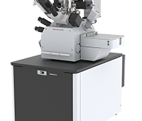
 Thermo Fisher Scientific has introduced two breakthrough imaging filters, the Thermo Scientific Selectris Imaging Filter and the Thermo Scientific Selectris X Imaging Filter, taking cryo-electron microscopy (cryo-EM) to a new level, making it possible to view proteins at true atomic resolution.
Combined with a Thermo Scientific Krios or Glacios Cryo-Transmission Electron Microscope (TEM), the Selectris filters allow biologists to accelerate their structural analysis research by viewing protein structures in unprecedented detail and in less time than was previously possible.
“Cryo-EM has already been a game changer, and our Selectris filters solidify this technique as a ‘must have’ for mapping 3D protein structures,” said Trisha Rice, vice president and general manager of life sciences at Thermo Fisher Scientific. “With a more precise understanding of the connections between proteins and disease, researchers can now speed the path to disease understanding and treatment.”
In a study published on bioRxiv, a popular pre-print server for biology, structural biologists used a Krios equipped with a Selectris prototype to study protein structures at never-before-achieved resolutions. Researchers from the Medical Research Council Laboratory of Molecular Biology in Cambridge, UK, obtained a 1.2 ångström resolution structure of the iron-storing protein apoferritin. They also achieved a 1.7 ångström resolution of the GABA Type A receptor-associated protein, with an even sharper resolution in key parts of the protein. This human membrane protein has target sites for general anesthetics, benzodiazepines, barbiturates and neuroactive steroids, making it very relevant to drug discovery.
“The image contrast is vastly improved with the Selectris, enabling us to view precise molecular details. This could, for example, help us determine where a drug would displace water molecules,” said Radu Aricescu, a study co-author and structural biologist at the Medical Research Council Laboratory of Molecular Biology in Cambridge, UK. “This has positive implications for structure-based drug design, potentially leading to better medications with fewer side effects.”
The Selectris X was designed for researchers seeking industry-leading optical performance, while the Selectris is ideal for those who require maximum efficiency and throughput. Both filters offer an unmatched combination of stability and contrast, allowing researchers to produce high quality data, generate fast results, and push the boundaries of resolution to improve single particle analysis and cryo-electron tomography results. The stability of the filters also reduces the need for user intervention and tuning, improving experiment productivity.
Both the Selectris X and Selectris imaging filters are available with the Krios and Glacios. For more information, visit ter.li/Selectris.


Tell Us What You Think!