A research collaboration between Southwest Research Institute (SwRI) and the University of Texas at San Antonio (UTSA) uses machine vision algorithms to track how brain cells, or neurons, develop over time. The team aims to better understand brain cell behavior and identify new therapies for neurological disorders by monitoring how new neurons grow and integrate into existing networks.
Innovative method for monitoring neural growth
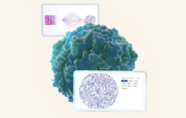
UTSA researchers have cultivated human stem cells into living neuronal networks that mimic areas of human brain tissue associated with sleep, temperature regulation, and mood. Using confocal microscopy, they recorded time-lapse images of these maturing cell networks. SwRI then developed a U-Net machine-learning algorithm to recognize and analyze key neuron structures from those images.
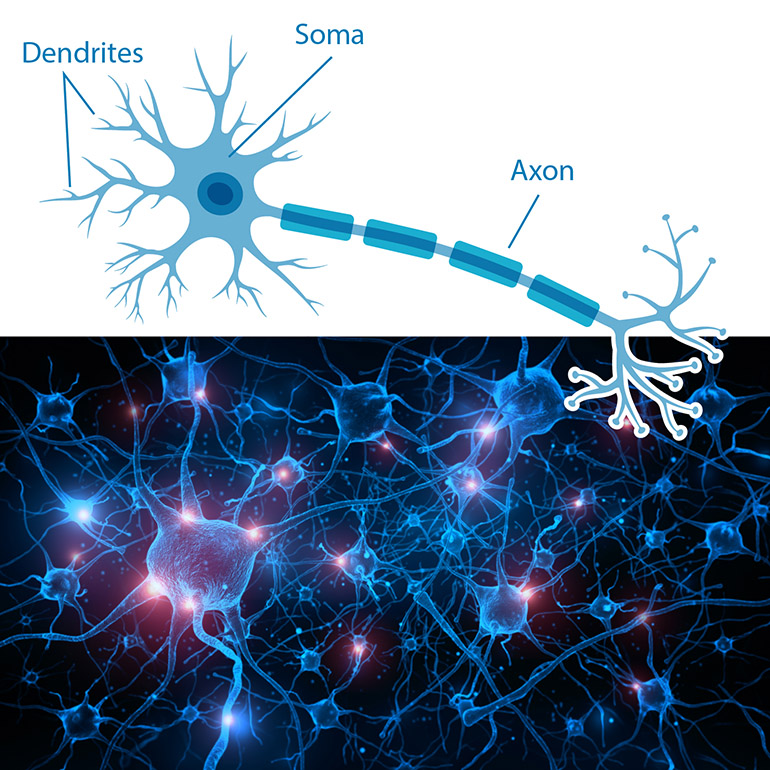
A team of researchers at Southwest Research Institute (SwRI) and The University of Texas at San Antonio (UTSA) developed a computational method that improves the tracking of neurogenesis as neurons grow within neuronal networks. A neuron has three main parts — a soma, dendrites and axon (top illustration) — to send and receive signals to other cells in a neuronal network, in a human brain (bottom illustration). Courtesy of SwRI
Conventional methods for studying neurons typically rely on fixed-tissue imaging, which limits insight into how cells behave in real time. In contrast, SwRI’s computational approach follows unlabeled cells and fine structures in dense, living cultures, allowing researchers to capture cellular activity over extended periods. The San Antonio Medical Foundation awarded a $200,000 grant to support this project.
“The research results are a significant step toward automatically classifying the health of growing neuronal networks. The algorithm can help study various neurological diseases and assist in the development and testing of associated therapies.” — Dr. Courtney Rouse, a SwRI computer scientist who led the project
Potential outcomes of the research
- Identifying potential therapies for neurological disorders
Scientists may pinpoint promising drug candidates that enhance or inhibit neural growth by observing how individual neurons respond to different treatments or conditions. - Accelerating drug discovery
The automated method could serve as a screening tool, enabling researchers to evaluate the effects of various compounds on neuron health and connectivity rapidly. - Improving understanding of brain development
Tracking live cells in maturing networks may reveal new information about the processes that underlie normal brain function and point to early interventions for neurological conditions. - Refining AI models for biomedical research
The techniques applied by SwRI and UTSA could be adapted for other cell types and tissues, providing a platform for more sophisticated biomedical data analysis. - Guiding future experiments on environmental and physiological stressors
Researchers plan to test neurons exposed to low oxygen levels, circadian rhythm disruptions, and other stresses, observing changes in neuron growth and function to gain further insights into tissue health.
Tracking and analysis
The new tracking algorithm focuses on the “soma” — the cell body where the nucleus resides — because each neuron has exactly one. Attaching a reference point (or tracking number) to each soma allows the system to follow these cells from image to image, achieving high precision and recall rates. The algorithm can also detect dendrites, though their smaller size presents additional challenges.
Future research
Next steps include establishing how dendrites connect to somas and correlating electrical activity with overall tissue health. Dr. Amina Qutub’s UTSA lab, which helped develop the experimental models, plans to integrate SwRI’s cell-tracking techniques into enhanced artificial intelligence methods. This progression may eventually enable more detailed monitoring of neuronal function in conditions resembling real-world stresses and environments.
The SwRI-UTSA partnership highlights the growing role of computational tools in medical research, with the aim of fostering more accurate diagnoses, treatments, and preventive strategies for disorders that affect the nervous system. Researchers say the methods honed in this project will help medical professionals make more informed decisions when dealing with complex data about brain cell health.