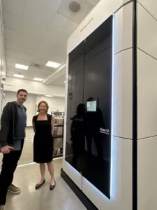
The author (left) standing next to Melanie Adams-Cioaba (right), Thermo Fisher’s senior director and general manager for life sciences electron microscopy, alongside the Krios G4 cryo-TEM at the company’s South San Francisco drug discovery center, which opened in March 2026.
The resolution revolution in cryo-electron microscopy had reached broad recognition by 2015, when Nature Methods hailed it the method of the year and declared an end to “blob-ology,” a term structural biologist Helen Saibil recalls crystallographers once used to dismiss EM’s then low-resolution imaging. In the decade since, pharma companies have moved from cautious exploration to building cryo-EM into their core discovery platforms, and that maturation is part of what led Thermo Fisher Scientific to open a Cryo-EM Drug Discovery Center in South San Francisco in March 2026, giving U.S. pharma and biotech companies a counterpart to the UK Pharmaceutical Cryo-EM Consortium in Cambridge, which the company launched in 2016.
The UK consortium served as the proof of concept. Adams-Cioaba said every UK consortium member went on to invest in house, some purchasing multiple instruments, while maintaining the consortium relationship for resiliency and access to Thermo Fisher’s applications road maps. U.S. customers, meanwhile, kept asking why the company hadn’t done the same stateside.
“When the UK site was launched, and being on the other side of the table, customers asked: Why not in the U.S.?” said Melanie Adams-Cioaba, Thermo Fisher’s senior director and general manager for life sciences electron microscopy, during a tour of the new facility.
Shared access before the capital commitment
In its announcement, Thermo Fisher framed the center around speed. Structure-guided drug programs, the company says, reach key preclinical milestones in about half the time and at roughly half the cost of industry averages, with more than twice the rate of clinical success. South San Francisco, in the middle of the Bay Area biotech corridor, puts the facility within commuting distance of a dense cluster of potential users, but the center also serves customers remotely. “They’re able to send us samples, we get the system up and ready, load the samples in, and they’re able to access remotely and drive their own data acquisition and live stream data back to them,” Adams-Cioaba said. “It could be in the basement, it can be across the street, or it can be anywhere else in the world.”
Adams-Cioaba drew a line between the center and the growing number of contract research organizations offering cryo-EM services. “We’re not a fee-for-service site,” she said. “We’re not here to just intake samples and spit back structures.” The model is closer to a core lab, where member companies can collect their own data and control their own workflows.
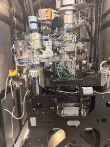
The interior of the Krios G4 cryo-TEM, showing the column and optical train behind the instrument’s outer housing.
To be sure, a high-end cryo-EM microscope like the Krios, with supporting infrastructure for sample prep, data processing and purpose-built facility construction, represents a significant capital outlay. Instruments are assembled to order, often with bespoke configurations. Meanwhile, facility work, including sample-prep space, data-handling infrastructure, vibration control and environmental mitigation, can add substantially to total deployment cost and complexity.
“You don’t go from one tool to two tools overnight,” Adams-Cioaba said. “You go from one tool to one and a quarter, one and a half, one and three quarters, and then your finance says, yes, you can buy another one.”
Inside the microscope room
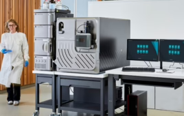
The center houses a Krios G4 cryo-TEM, which uses a 300 kV beam, a cold field emission gun, a Selectris X imaging filter and a Falcon 4i direct electron detector. Alongside the microscope sit Vitrobot systems for sample preparation.
Getting a sample into the Krios is a chain of precise steps that begins well before the microscope. Protein construct design, expression, purification and stabilization all determine whether the run produces usable data. The physical preparation involves applying a few microliters of purified protein to a TEM grid, a mesh smaller than a fingernail, then blotting away excess liquid and plunging the grid into liquid ethane for rapid vitrification. The sample stays at roughly minus 190 degrees Celsius from that point on. Ice thickness is critical.
A 24-hour data collection run on a target like a GPCR generates in the ballpark of 1 to 1.5 terabytes, depending on magnification. Thermo streams that data to remote customers in near-real time over a dedicated Ethernet connection.
From crystallography to cryo-EM
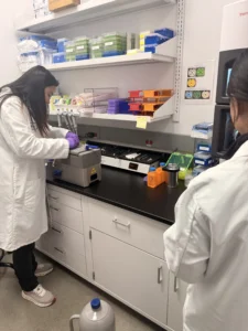
Researchers prepare samples at the Vitrobot vitrification station inside Thermo Fisher’s new Cryo-EM Drug Discovery Center in South San Francisco. Sample preparation, including protein purification and grid preparation, determines whether a data collection run yields usable structural data.
Structure-based drug design predates cryo-EM by decades, and X-ray crystallography, for all its productivity, requires coaxing a protein into a large, ordered crystal, a process that is unpredictable, can take years and is impractical for many complex or membrane-bound targets. Adams-Cioaba put the challenge in practical terms: “Imagine that you have to design a plug, and it has to fit your wall socket perfectly. Nobody’s given you a picture of the wall socket, and nobody’s given you the measured dimensions of what you’re trying to get.”
Cryo-EM bypasses crystallization by preserving purified particles in vitreous ice after rapid plunge-freezing, and in doing so provides that picture. Because the technique captures ensembles of particles in near-native states outside a crystal lattice, researchers can sort the same molecule into open, closed and intermediate conformations. Adams-Cioaba described it as capturing snapshots of proteins as they “dance,” pulling multiple structures from a single dataset. This capability allows scientists to computationally reconstruct molecular motion by pulling multiple structures from a single dataset.
“Our ability to let them move and let them dance, and just capture them in their native state through vitrification, and then load them into these high-end tools, essentially we’ve expanded the universe of what’s possible in terms of the drug targets that we can see and design specifically for,” she said.
Membrane proteins, including GPCRs and ion channels, account for a large share of drug targets, and many resist crystallization. Cryo-EM’s ability to characterize them structurally has expanded the druggable universe, and the clinical returns are starting to show. Adams-Cioaba cited Thermo Fisher figures from publicly disclosed structure-guided programs: rational design approaches show roughly 2.5 times the likelihood of clinical success compared with an industry-average rate of about 10%, while cutting concept-to-clinic time, cost and risk by about half. A 2025 benchmark of 18 leading pharma companies put the average Phase I-to-approval probability at 14.3%, with company-level results ranging from 8 to 23%.
Computational tools like AlphaFold have accelerated the prediction side of structural biology, but Adams-Cioaba noted that general models reach a ceiling at the resolution that matters for drug design. “What we can’t do is know the difference of an angstrom in an interaction between a small molecule and its target protein,” she said. “That can fundamentally change the drug-like properties and the effectiveness of that therapeutic.”