The LCLS Coherent X-ray Imaging Experimental Station. Credit: Nathan Taylor, SLAC National Accelerator Laboratory
The world at the atomic scale is never at rest, with particles moving so quickly and molecular bonds changing so rapidly that we have been unable to capture their motion directly until now. Previously, we’ve had to rely on static pictures of the molecular world (using X-ray synchrotrons or electron microscopes), or infer dynamic behavior from spectroscopic signatures (using short-pulse optical lasers). All that changed in 2009, when the world’s first X-ray free-electron laser (XFEL) was successfully commissioned. The field of ultrafast X-ray science took a huge leap forward – with a source that was billions of times brighter than anything that came before, delivering bursts of X-rays on timescales that are many orders of magnitude shorter – reaching the femtosecond domain.
A flash of light as short as this can freeze the motion of atoms in molecules, allowing us to make slow-motion movies of how nature works. A femtosecond (a millionth of a billionth of a second) is at the fundamental scale of atomic and molecular physics, and so underpins the initiating events of the chemical, material, and biological processes that make up our world.
The concept of a free-electron laser was introduced by John Madey at Stanford in 1971, in which the passage of an electron beam through a series of magnets causes the emission of photons, which ultimately can interact to create a coherent burst of light. Later, in the 1990s, Claudio Pellegrini and collaborators proposed to extend free-electron lasers to the X-ray regime – a hugely ambitious concept that was ultimately proven with the construction of the Linac Coherent Light Source (LCLS) by the U.S. Department of Energy (DOE) at the SLAC National Accelerator Laboratory.
This new source combines three critical features. First, the wavelength of the X-ray light is at the Angstrom scale, and tunable, so that the individual atoms in a molecule can be imaged. Second, these X-rays are delivered on a short enough timescale to freeze the motion of atoms in molecules, capture the initiating events of molecular bond formation and the dynamics of electrons as they orbit an atom or carry charge around a molecule. Third, over a trillion X-rays are delivered in each pulse, resulting in a source that is so incredibly bright it can deliver precise information from just a single pulse. With over a hundred pulses per second, movies can be made of how atomic and molecular systems evolve – allowing unprecedented insight into fields as diverse as chemical catalysis, structural biology, quantum materials science, and the physics of planetary formation.
What underpins these fields is the need to make direct observations of fundamental charge, spin, and orbital or lattice dynamics on natural timescales – put simply, to see the motion of electrons and ions as they respond to their environment or to external stimuli.
From a capability point of view, the progress in XFEL performance has been dramatic, creating precision tools with unprecedented peak intensity and time-averaged brightness. In less than a decade, the X-ray pulse duration has been shortened from over 100 femtoseconds to 5 fs (and likely 0.5 fs later this year); full polarization control has been introduced, so we can see chiral molecules that are important for many pharmaceutical drugs; and a wide array of dual-pulse options have been developed that provide the ability to drive a system and monitor its response on timescales that range from femtoseconds to microseconds.
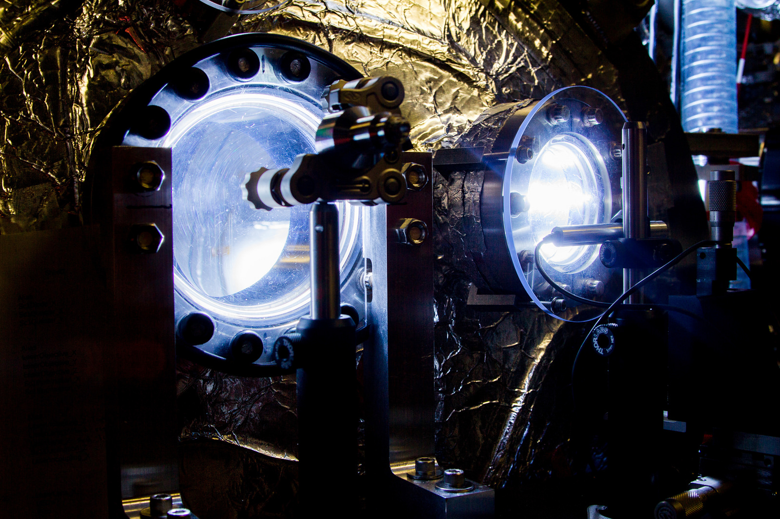
This is an illustration of an electron beam traveling through a niobium cavity – a key component of SLAC’s future LCLS-II X-ray laser. Kept at minus 456 degrees Fahrenheit, a temperature at which niobium conducts electricity without losses, these cavities will power a highly energetic electron beam that will create up to 1 million X-ray flashes per second – more than any other current or planned X-ray laser. Credit: SLAC National Accelerator Laboratory
Looking forward
The pace of progress is set to further accelerate over the next few years. The first X-ray laser, LCLS, delivered 120 pulses per second, followed by SACLA in Japan at 60 per second. A new facility, the European-XFEL based in Hamburg, Germany turned on in mid-2017, delivering pulses at 27,000 per second. And now an additional $1 billion is being invested by the DOE to create the LCLS-II upgrade that will provide up to a million pulses per second by 2020. This will be transformative, allowing the study of real-world systems that are simply inaccessible today, including statistical fluctuations and heterogeneous materials, rather than idealized samples.
Normally when a field advances, the performance of the system increases incrementally, or sometimes by a factor of 10 or so. Here, the field had to cope with a factor of a billion increase in capability – requiring a completely new approach to the measurements, and innovation in almost all aspects of the technology.
The incredible efforts in delivering this novel X-ray source are thus only part of the story. Precision science requires the integration of new instrumentation and measurement techniques that can take advantage of the characteristics of the new source, and so provide quantitative information. This in turn requires detectors that can sense photons individually or by the thousands and operate at the same repetition rate as the source; optics that can handle the intense X-ray power; delivery of a continuous stream of samples for the X-rays to probe; and data acquisition systems that can cope with unprecedented rates.
Applications across industries
The degree to which this has been achieved is astonishing, and has touched a very large number of fields. In chemical science, the ultrashort bursts of XFEL light have been used to capture the birth of a chemical bond and follow the ultrafast dynamics of catalytic activity as gas is passed over a metal surface – allowing insight into the fundamental reactions that drive major industrial processes.
Similarly, the brightness of the beam has been used to create detailed “molecular movies” that for the first time track the opening of a ring molecule after a bond is broken by a burst of light. This ability to watch molecules evolve their geometrical structure in real time removes the great uncertainties associated with only being able to see the initial and final stages of structure formation and having to rely on complex numerical models.
Interestingly, this approach has had great impact in the complex field of structural biology and the associated problem of drug discovery for improved medicines. In that area, LCLS has revolutionized the study of membrane proteins by allowing a new technique known as “diffraction before destruction” that can measure the atomic structure of very delicate samples. It has also allowed observations of how molecular machines work in living systems with sub-picosecond precision. Applications have ranged from investigating neurotransmitters connected with the study of Alzheimer’s disease, to finding weaknesses in the parasite that causes African sleeping sickness, to gaining a new understanding of how the body can regulate blood pressure and anxiety. Moving to the atomic scale, LCLS has provided the first direct evidence of superfluidity in nanometer-sized quantum systems, and has imaged the process of electron charge transfer to better understand how to harness photosynthesis for energy generation.
With the advent of the new XFEL sources in Japan, Germany, the United States, Switzerland and the Republic of Korea, this field is set to expand into many new research areas. Much work is currently underway to define the most compelling science opportunities, and thus guide the direction of facility development. But the most impactful science may well come from fields that have never previously used X-ray sources or been able to peer into the ultrafast world.