Research & Development World

  • R&D World Home
  • Topics
    • Aerospace
    • Automotive
    • Biotech
    • Careers
    • Chemistry
    • Environment
    • Energy
    • Life Science
    • Material Science
    • R&D Management
    • Physics
  • Technology
    • 3D Printing
    • A.I./Robotics
    • Software
    • Battery Technology
    • Controlled Environments
      • Cleanrooms
      • Graphene
      • Lasers
      • Regulations/Standards
      • Sensors
    • Imaging
    • Nanotechnology
    • Scientific Computing
      • Big Data
      • HPC/Supercomputing
      • Informatics
      • Security
    • Semiconductors
  • R&D Market Pulse
  • R&D 100
    • Call for Nominations: The 2025 R&D 100 Awards
    • R&D 100 Awards Event
    • R&D 100 Submissions
    • Winner Archive
    • Explore the 2024 R&D 100 award winners and finalists
  • Resources
    • Research Reports
    • Digital Issues
    • Educational Assets
    • R&D Index
    • Subscribe
    • Video
    • Webinars
  • Global Funding Forecast
  • Top Labs
  • Advertise
  • SUBSCRIBE

New cryo-EM technology at St. Jude Children’s Research Hospital pushes boundaries of molecular imaging

By Brian Buntz | September 13, 2024

A media release and Scientific Report image of Tharun Chand Pottangadi and Elizabeth Kellogg.

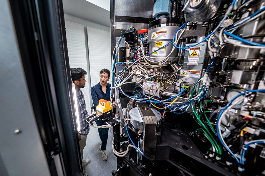
Elizabeth Kellogg, Ph.D. (right) and Tharun Chand Pottangadi, researchers at St. Jude Children’s Research Hospital, standing near the Krios G4 Cryo-TEM microscope. [St. Jude Children’s Research Hospital.]

For decades, structural biologists have sought to map the structures of proteins and other macromolecules within cells. Cryo-electron microscopy (cryo-EM), whose early development traces back to the 1970s, emerged as a promising technique for visualizing cells in their natural, hydrated state. But the technology’s initial inability to produce sharp images was a stumbling block in the early days. Early cryo-electron microscopes, while still a breakthrough, produced images so blurry that the field earned the self-deprecating nickname “blob-ology.” It wasn’t until around 2012 that advances in detectors and image processing software propelled cryo-EM to widespread popularity and recognition, ushering in a “resolution revolution,” as Science put it in 2017.

Pushing resolution boundaries

Now, Liz Kellogg, Ph.D., Associate Member in the Department of Structural Biology at St. Jude Children’s Research Hospital, is among a select group of researchers with access to a new cryo-EM instrument capable of pushing the resolution boundaries further. “In terms of cryo-EM in the U.S., there might be two other institutions that have similar setups,” Kellogg adds. “And what’s so special about this setup is that it is able to generate atomic-resolution structures and will be great to push our cryo-ET efforts.” [Learn more about St. Jude’s gene editing research here.]

Cryo-electron tomography (cryo-ET) is a specialized form of cryo-electron microscopy that allows scientists to visualize the 3D structure of biological samples, like cells or tissues, in their natural environment. The technique involves rapidly freezing the sample to preserve its structure and then imaging it from multiple angles via an electron microscope. These images are then combined computationally to create a detailed 3D model.

The new cryo-EM microscope, part of the St. Jude Cryo-Electron Microscopy and Tomography Center, is a Titan Krios G4 equipped with a cold FEG electron source, a SelectrisX energy filter, and a Falcon 4i camera. The cold FEG electron source optimizes beam coherence and brightness, enabling exceptionally high-resolution imaging. The energy filter improves enhanced contrast and the signal-to-noise ratio in acquired images. The Falcon 4i camera supports rapid, high-sensitivity electron detection and crisp image quality at both high and low magnifications.

“All of these elements come together to produce high-quality, detailed images,” Kellogg explained. “This is a highly customized setup that represents a significant investment.”

A media release and Scientific Report image of Elizabeth Kellogg and Tharun Chand Pottangadi. - Camera Settings: ILCE-9M2, 12mm, ISO 1000, 1/50, f/3.2, Fri, 04-19-2024 at 10:17. v.12.01.23

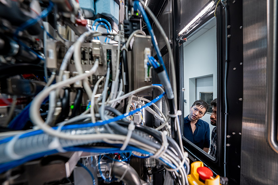
Elizabeth Kellogg, Ph.D. (left) and Tharun Chand Pottangadi. [St. Jude Children’s Research Hospital]

Down to the atomic level

“This new microscope can achieve resolutions down to 1.2 angstroms (Å),” Kellogg said. To put that into perspective, that’s essentially the resolution scale of X-ray crystallography, a technique that enables scientists to see the separation between atoms.

“At this level of resolution, we can see the atomic arrangements that allow molecules to execute their biological functions,” Kellogg said. “We can also observe exactly how a small molecule binds to specific sites in a macromolecule, which is essential for drug discovery.”

Inside the advanced electron microscopy facility at St. Jude

The Titan Krios G4 microscope’s ability to achieve resolutions as high as 1.2 angstroms opens up new possibilities for understanding the workings of biological systems. “To understand a biological process in detail, we need to be able to see how it happens with precision” Kellogg said. Researchers are now able to see how individual atoms are arranged within a protein, how a water molecule nestles between a protein and DNA, and how a drug molecule fits into its target binding pocket. “I know a lot of people at St. Jude are interested in looking at drug interactions,” Kellogg said. “And to drive drug discovery, you really need that level of precision to observe how a small molecule binds to its target.”

At St. Jude, the availability of powerful tools such as the new cryo-EM microscope is also a factor attracting other researchers aimed to help tackle severe pediatric diseases. “I think we have a absolutely fantastic group of talented, passionate scientists,” Kellogg explained. “And everybody is motivated by our shared mission to cure fatal childhood diseases.”

Comments

  1. Premagiri e says

    November 11, 2024 at 12:29 am

    Really wonderful work
    convey my regards and appreciation to team members Ms.Elizabth kellogg
    and Mr.Tharunchand pottangadi
    wish you all the best

  2. Jessy Ashok says

    November 12, 2024 at 3:12 am

    Great work Tarun , really proud of you. Wish you all the best in all your future endeavours

  3. JEENA JAYARAM says

    November 12, 2024 at 8:29 pm

    Really proud of you Tarun..Amazing work. Wish you all the best

Related Articles Read More >

How materials scientists are using (S)TEM to better understand our world on the atomic scale 
Chromium X (GEM-X Flex) instrument on the bench
10x CTO on Chan Zuckerberg, Arc Institute ties and industrializing single-cell biology
Thermo Fisher’s new Orbitrap Excedion Pro targets complex biotherapeutics for drug development
Peak Scientific trims lab-gas footprint with new Intura series at ASMS 2025
rd newsletter
EXPAND YOUR KNOWLEDGE AND STAY CONNECTED
Get the latest info on technologies, trends, and strategies in Research & Development.
RD 25 Power Index

R&D World Digital Issues

Fall 2024 issue

Browse the most current issue of R&D World and back issues in an easy to use high quality format. Clip, share and download with the leading R&D magazine today.

Research & Development World
  • Subscribe to R&D World Magazine
  • Enews Sign Up
  • Contact Us
  • About Us
  • Drug Discovery & Development
  • Pharmaceutical Processing
  • Global Funding Forecast

Copyright © 2025 WTWH Media LLC. All Rights Reserved. The material on this site may not be reproduced, distributed, transmitted, cached or otherwise used, except with the prior written permission of WTWH Media
Privacy Policy | Advertising | About Us

Search R&D World

  • R&D World Home
  • Topics
    • Aerospace
    • Automotive
    • Biotech
    • Careers
    • Chemistry
    • Environment
    • Energy
    • Life Science
    • Material Science
    • R&D Management
    • Physics
  • Technology
    • 3D Printing
    • A.I./Robotics
    • Software
    • Battery Technology
    • Controlled Environments
      • Cleanrooms
      • Graphene
      • Lasers
      • Regulations/Standards
      • Sensors
    • Imaging
    • Nanotechnology
    • Scientific Computing
      • Big Data
      • HPC/Supercomputing
      • Informatics
      • Security
    • Semiconductors
  • R&D Market Pulse
  • R&D 100
    • Call for Nominations: The 2025 R&D 100 Awards
    • R&D 100 Awards Event
    • R&D 100 Submissions
    • Winner Archive
    • Explore the 2024 R&D 100 award winners and finalists
  • Resources
    • Research Reports
    • Digital Issues
    • Educational Assets
    • R&D Index
    • Subscribe
    • Video
    • Webinars
  • Global Funding Forecast
  • Top Labs
  • Advertise
  • SUBSCRIBE