
Today, tracking the development of individual cells and spotting the associated factors under the microscope is nothing unusual. However, impairments like shadows or changes in the background complicate the interpretation of data. Now, researchers at the Technical University of Munich (TUM) and the Helmholtz Zentrum München have developed a software that corrects images to make…
How Next-Gen Thermal Imaging Can Help Keep Firefighters Safe
Thermal Imaging Cameras (TICs) are proven lifesavers for fire crews with an established track record in all phases of fire size-up, attack and, overhaul. Not only can they help firefighters find their way through thick smoke in unfamiliar surroundings, TICs can also help crews determine the center of fire activity, locate victims and other firefighters,…
Combining MRI and Optical Microscopy Promising for Brain Research

Functional magnetic resonance imaging reveals changes in blood-oxygen levels in different parts of the brain, but the data show nothing about what is actually happening in and between brain cells, information needed to better understand brain circuitry and function. “We really have no clear understanding of what cellular processes cause the MRI signal and are…
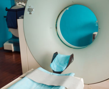
Medical Gamma-Ray Camera is now Palm-Sized

As represented by conventional radiograph, radiological images provide only black and white figures in 2D space. The situation is basically the same for Single photon emission tomography (SPECT) and positron emission tomography (PET), which are the two most common molecular imaging techniques used in nuclear medicine. PET is used especially for early cancer and Alzheimer’s…
Laser, Sound Waves Provide Live Views of Organs in Action

Biomedical engineers are now able to take a live, holistic look at the inner workings of a small animal with enough resolution to see active organs, flowing blood, circulating melanoma cells and firing neural networks. The technique dubbed “single-impulse photoacoustic computed tomography (SIP-PACT)” uses the best of both light and ultrasound to peer inside living…
‘Spectral Fingerprinting’ Sees Through Concrete to Detect Early Corrosion

When you suffer a fall, an on-the-field collision or some other traumatic blow, the first thing the doctor will do is take an X-ray, CT scan or MRI to determine if anything has been damaged internally. Researchers at the National Institute of Standards and Technology (NIST) are using the same principle, but in a more powerful form,…
Cameras Reveal Images Hidden to the Naked Eye

EPFL researchers took advantage of the limits of human vision to hide an image in a video. The image is invisible to the human eye, but not to a camera. Human visual perception works well and is very effective at seeing what’s important to us. But our eyes are not capable of averaging video images…
Technique Makes More Efficient, Independent Holograms

Not far from where Edwin Land — the inventor of the Polaroid camera — made his pioneering discoveries about polarized light, researchers from the Harvard John A. Paulson School of Engineering and Applied Sciences (SEAS) are continuing to unlock the power of polarization. Recently, a team of researchers led by Federico Capasso, the Robert L.…
New Detection System Makes Molecules More Visible in the Body

IBM, Medymatch Partner on Imaging Technology for Brain Bleeding
Imaging at the Speed of Light
Tiny micro- and nanoscale structures within a material’s surface are invisible to the naked eye, but play a big role in determining a material’s physical, chemical, and biomedical properties. Over the past few years, Chunlei Guo and his research team at the University of Rochester have found ways to manipulate those structures by irradiating laser…
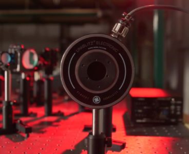
Imaging High Explosive Detonators
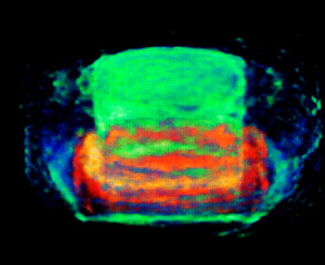
Lawrence Livermore National Laboratory (LLNL) scientists and collaborators at Los Alamos National Laboratory(link is external) (LANL) for the first time have taken 3D snapshots of operating high explosive detonators. Scientists from LLNL, Los Alamos and National Security Technologies, LLC (link is external)(NSTech) combined state-of-the-art imaging capabilities with computed tomographic reconstruction (X-ray cross sectional imaging) in experiments performed at the Argonne National…
Breakthrough in Live Coral Imaging

Super Resolution Imaging Helps Determine a Stem Cell’s Future

Laser-Generated Bubbles Create 3-D Images in Liquid

Researchers have developed a completely new type of display that creates 3D images by using a laser to form tiny bubbles inside a liquid “screen.” Instead of rendering a 3D scene on a flat surface, the display itself is three-dimensional, a property known as volumetric. This allows viewers to see a 3D image in the…
Why Medical Technology Often Doesn’t Make It From Drawing Board to Hospital
Laser-Based Camera Improves View of Carotid Artery
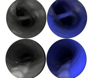
Strokes and heart attacks often strike without warning. But, a unique application of a medical camera could one day help physicians know who is at risk for a cardiovascular event by providing a better view of potential problem areas. A new paper in Nature Biomedical Engineering reports proof-of-concept results for this new imaging platform for atherosclerosis. “The…
Terahertz Wave Microchip Furthers Spectroscopy, Security, Medical Imaging
A new method to harvest terahertz waves— electromagnetic pulses which last one millionth of a millionth of a second—may be the key to advances in medical imaging, communications and drug development. While utilizing these pulses previously required elaborate and expensive equipment, a research team at Princeton University believes they have drastically streamlined the process by…
Novel Liquid Crystal Could Triple Sharpness of Today’s Televisions

An international team of researchers has developed a new blue-phase liquid crystal that could enable televisions, computer screens and other displays that pack more pixels into the same space while also reducing the power needed to run the device. The new liquid crystal is optimized for field-sequential color liquid crystal displays (LCDs), a promising technology…
Imaging Technique Measures Tumor Stiffness to Aid Surgical Planning
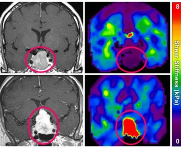
Important steps in planning tumor surgery include identifying borders between tumor and healthy tissue and assessing the tumor stiffness, e.g. hard and calcified or soft and pliant. For decades, tumors near the surface of the body have been evaluated for stiffness by simple palpation—the physician pressing on the tissue. Because tumors within the skull cannot…
X-Ray Imaging at Argonne Captures Material Defect Process

From blacksmiths forging iron to artisans blowing glass, humans have for centuries been changing the properties of materials to build better tools – from iron horseshoes and swords to glass jars and medicine vials. In modern life, new materials are created to improve today’s items, such as stronger steel for skyscrapers and more reliable semiconductors…
New Technology Enables 5D Imaging in Live Animals, Humans

A new image analysis technique makes it easier for scientists to quickly find and track important biological molecules including tell-tale signs of disease. Called “Hyper-Spectral Phasor” analysis, or HySP, it could even be useful for diagnosing and monitoring diseases by using cell phone images. It is much faster and far less expensive than current techniques.…
Chemistry on the Edge

Defects and jagged surfaces at the edges of nanosized platinum and gold particles are key hot spots for chemical reactivity, a team of researchers working at the Department of Energy’s Lawrence Berkeley National Laboratory (Berkeley Lab) and the Hebrew University of Jerusalem in Israel confirmed with a unique infrared probe. Experiments like this should help…
Optical Device Offers Widest Real-Time Views of Vast Regions of the Sun

A groundbreaking new optical device, developed at NJIT’s Big Bear Solar Observatory (BBSO) to correct images of the Sun distorted by multiple layers of atmospheric turbulence, is providing scientists with the most precisely detailed, real-time pictures to date of solar activity occurring across vast stretches of the star’s surface. The observatory’s 1.6-meter New Solar Telescope…
New Microscope Chemically Identifies Micron-Sized Particles

Researchers have developed a microscope that can chemically identify individual micron-sized particles. The new approach could one day be used in airports or other high-security venues as a highly sensitive and low-cost way to rapidly screen people for microscopic amounts of potentially dangerous materials. In the journal Optics Letters, from The Optical Society (OSA), researchers from the Massachusetts…