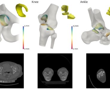
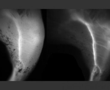
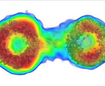
An algorithm to monitor the joints of patients with arthritis, which could change the way that the severity of the condition is assessed, has been developed by a team of engineers, physicians and radiologists led by the University of Cambridge. The technique, which detects tiny changes in arthritic joints, could enable greater understanding of how…
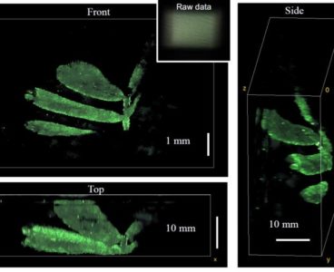
3D Imaging and Computer Modeling Capture Breast Duct Development
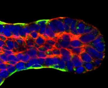
Working with hundreds of time-lapse videos of mouse tissue, a team of biologists joined up with civil engineers to create what is believed to be the first 3D computer model to show precisely how the tiny tubes that funnel milk through the breasts of mammals form. A report on the model was published April 9 in Developmental Cell.…
Dolphin Echolocation Discovery Could Improve Ultrasound Technology

Diamond ‘Spin-Off’ Tech Could Lead to Low-Cost Medical Imaging and Drug Discovery Tools
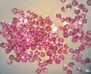
It may sound contradictory, but diamonds are the key to a new technique that could provide a very-low-cost alternative to multimillion-dollar medical imaging and drug-discovery devices. An international team led by scientists at the Department of Energy’s Lawrence Berkeley National Laboratory (Berkeley Lab) and UC Berkeley discovered how to exploit defects in nanoscale and microscale…
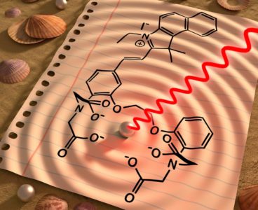
X-rays from Tabletop Lasers Allows Scientists to Peer Through the ‘Water Window’
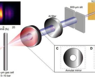
Studying the fleeting actions of electrons in organic materials will now be much easier, thanks to a new method for generating fast X-rays. The technique means advanced measurements of fast reactions will now be possible in physics labs around the world, without having to wait to use expensive and scarce equipment. It could be used,…
MRI Glove Gives Doctors a Deeper Look at the Hand
A new kind of MRI component in the shape of a glove delivers the first clear images of bones, tendons and ligaments moving together, a new study finds. Led by NYU School of Medicine and just published in Nature Biomedical Engineering, the study shows how a new MRI element design woven into garment-like detectors can capture…
X-Ray Laser Opens New View on Alzheimer’s Proteins

Using Infrared Light, New Pill Could More Accurately Detect Breast Cancer Tumors
Calcium-Based MRI Sensor Enables More Sensitive Brain Imaging
New Way of Producing Intense Radiation Could Offer Less Harmful Alternative to X-Rays
Improving Mid-Infrared Imaging and Sensing

New Imaging System Makes Back Surgery Safer, Faster and Less Expensive

Fluorescent Dye Could Enable Sharper Biological Imaging
Fluorescence imaging is widely used for visualizing biological tissues such as the back of the eye, where signs of macular degeneration can be detected. It is also commonly used to image blood vessels during reconstructive surgery, allowing surgeons to make sure the vessels are properly connected. For these procedures, as well as others now in…
New Technique Improves MRI Images
Powerful New Imaging Method Reveals in Detail How Particles Move in Solution
New research published in Nature Methods will dramatically improve how scientists “see inside” molecular structures in solution, allowing for much more precise ways to image data in various fields, from astronomy to drug discovery. The new method will allow for the visualization of many more biological molecules, providing critical information about what is inside molecules…
X-Rays Reveal ‘Handedness’ in Swirling Electric Vortices

Engineers Create Next-Generation Sensor for Multiple R&D Applications

Lensless Camera Captures Highly Detailed 3D Images
An experimental camera can produce extremely detailed images without even using a lens. Scientists from the University of California, Berkeley has created an easy-to-build lensless camera that produces 3D images from a single 2D image. “The DiffuserCam can, in a single shot, capture 3D information in a large volume with high resolution,” research team leader Laura Waller,…
MRI Technique Can Forecast Survival, Treatment Response in Patients with Brain Metastasis
New Hyperlens Can Capture Images of Living Cells
New hyperlenses are giving scientists the ability to capture images of living cells in great detail. A team from Vanderbilt University has created a new hyperlens that can resolve objects much smaller than the wavelength of light using hexagonal boron nitride (hBN)—a natural crystal with hyperlensing properties. Previously, the best resolution using hBN was an…
Molecular Beacon Signals Low Oxygen With Ultrasound
Areas of hypoxia, or low oxygen in tissue, are hallmarks of fast-growing cancers and of blockages or narrowing in blood vessels, such as stroke or peripheral artery disease. University of Illinois researchers have developed a way to find hypoxic spots noninvasively in real time. The researchers developed an oxygen-sensitive molecular beacon that emits ultrasound signals…
Seeing Through Walls of Unknown Materials
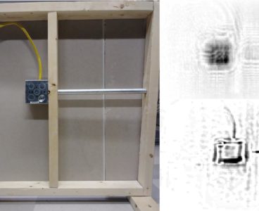
Researchers at Duke University have devised a way to see through walls using a narrow band of microwave frequencies without any advance knowledge of what the walls are made out of. Besides having obvious applications in the realm of security, the approach could lead to inexpensive devices to help construction workers easily locate conduits, pipes…
Compact Hyperspectral Imaging at Low Cost
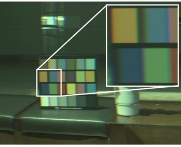
With hyperspectral imaging, photographers can obtain super fine detailed images, capturing the spectrum for each pixel in an image of a scene. This technology has wide reach and is being applied in fields such as military combat, astronomy, agriculture, biomedical imaging and geoscience. Scientists, for instance, rely on hyperspectral imaging to observe and analyze materials…
Ultrasound Imaging Needle to Transform Heart Surgery
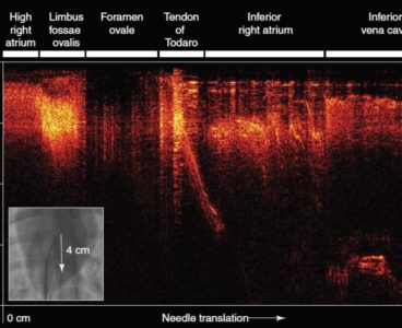
Heart tissue can be imaged in real-time during keyhole procedures using a new optical ultrasound needle developed by researchers at UCL and Queen Mary University of London (QMUL). The revolutionary technology has been successfully used for minimally invasive heart surgery in pigs, giving an unprecedented, high-resolution view of soft tissues up to 2.5 cm in…
Visible Signals From Brain and Heart
Key processes in the body are controlled by the concentration of calcium in and around cells. A team from the Technical University of Munich (TUM) and the Helmholtz Zentrum München have developed the first sensor molecule that is able to visualize calcium in living animals with the help of a radiation-free imaging technique known as…